. 2021 Jan 13;8(1):201785.
doi: 10.1098/rsos.201785. eCollection 2021 Jan.

Ion release from hydroxyapatite and substituted hydroxyapatites in different immersion liquids: in vitro experiments and theoretical modelling study

Affiliations

Ion release from hydroxyapatite and substituted hydroxyapatites in different immersion liquids: in vitro experiments and theoretical modelling study

Aurora Mocanu et al. R Soc Open Sci. .

Abstract

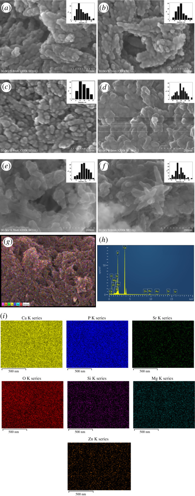
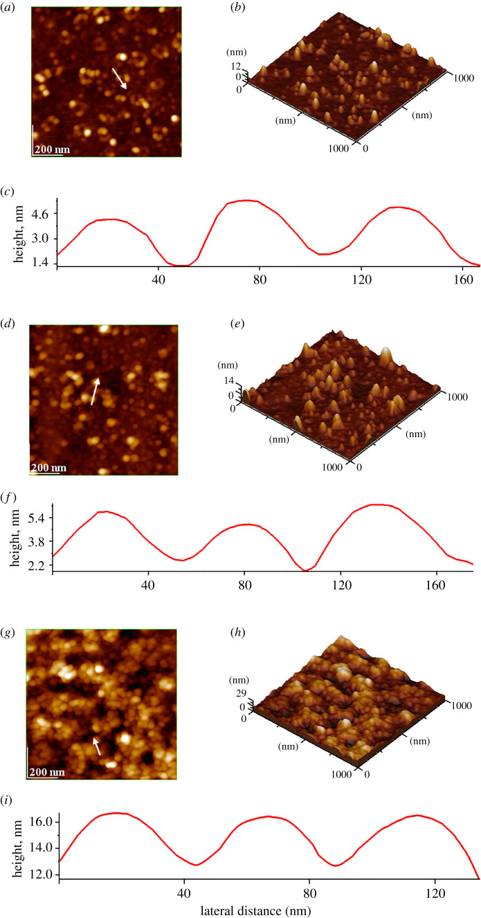
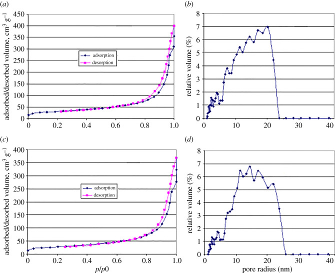
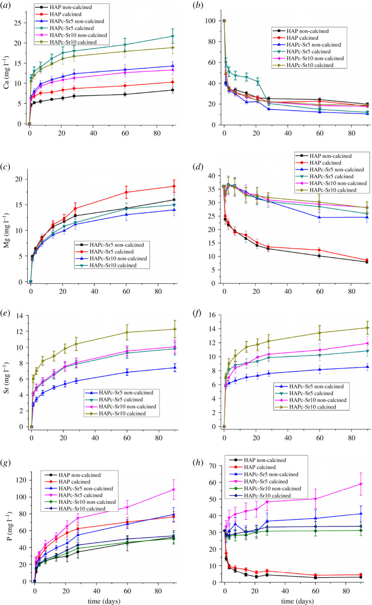
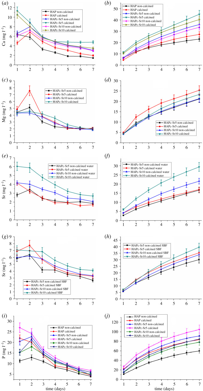
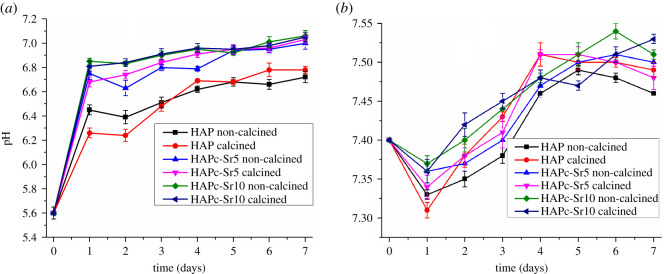
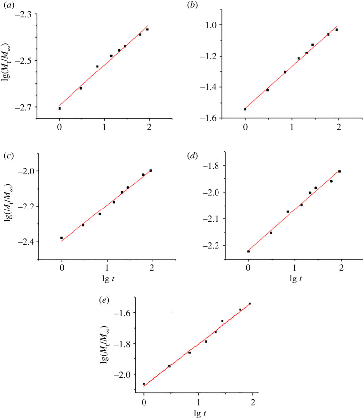
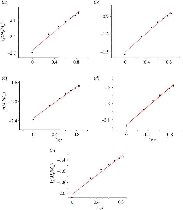
Multi-substituted hydroxyapatites (ms-HAPs) are currently gaining more consideration owing to their multifunctional properties and biomimetic structure, owning thus an enhanced biological potential in orthopaedic and dental applications. In this study, nano-hydroxyapatite (HAP) substituted with multiple cations (Sr2+, Mg2+ and Zn2+) for Ca2+ and anion ( Si O 4 4 - ) for P O 4 3 - and OH-, specifically HAPc-5%Sr and HAPc-10%Sr (where HAPc is HAP-1.5%Mg-0.2%Zn-0.2%Si), both lyophilized non-calcined and lyophilized calcined, were evaluated for their in vitro ions release. These nanomaterials were characterized by scanning electron microscopy, field emission-scanning electron microscopy and energy-dispersive X-ray, as well as by atomic force microscope images and by surface specific areas and porosity. Further, the release of cations and of phosphate anions were assessed from nano-HAP and ms-HAPs, both in water and in simulated body fluid, in static and simulated dynamic conditions, using inductively coupled plasma optical emission spectrometry. The release profiles were analysed and the influence of experimental conditions was determined for each of the six nanomaterials and for various periods of time. The pH of the samples soaked in the immersion liquids was also measured. The ion release mechanism was theoretically investigated using the Korsmeyer-Peppas model. The results indicated a mechanism principally based on diffusion and dissolution, with possible contribution of ion exchange. The surface of ms-HAP nanoparticles is more susceptible to dissolution into immersion liquids owing to the lattice strain provoked by simultaneous multi-substitution in HAP structure. According to the findings, it is rational to suggest that both materials HAPc-5%Sr and HAPc-10%Sr are bioactive and can be potential candidates in bone tissue regeneration.

Keywords: Ca; Korsmeyer–Peppas model; Mg and P release; Sr; multi-substituted hydroxyapatites; release mechanism.


Conflict of interest statement

we declare we have no competing interests.

Figures

Figure 1.
Figure 1.
SEM images and size distribution of nanoparticles for non-calcined samples: HAP (a), HAPc-5%Sr (b) and HAPc-10% Sr (c), and calcined samples: HAP (d), HAPc-5%Sr (e) and HAPc-10% Sr (f). The bars in the figures are 100 nm (ac) and 200 nm (df). Calcined HAPc-5%Sr: FE-SEM multi-colour distribution map of elements (g), EDX spectrum (h), distribution maps for each element (i). Gold appears in the EDX spectrum owing to coating of the nanomaterial with a gold layer for high imaging resolution.
Figure 2.
Figure 2.
AFM images: two-dimensional topographies (a,d,g) and three-dimensional images (b,e,h) of calcined HAP and ms-HAP samples as thin films, and cross-section profile (c,f,i) along the arrow given in two-dimensional topographies for HAP (ac), HAPc-5%Sr (df) and HAPc-10%Sr (gi); surface roughness (e.g. root mean square: RMS) on area and on profile are, respectively, for HAP, 4.58 nm (a) and 1.31 nm (c), for HAPc-5%Sr, 1.65 nm (d) and 1.29 nm (f) and for HAPc-10%Sr, 1.38 nm (g) and 1.29 nm (i).
Figure 3.
Figure 3.
Adsorption–desorption isotherms and pore radius distributions for non-calcined HAPc-5%Sr (a,b), and for calcined HAPc-5%Sr (c,d) samples.
Figure 4.
Figure 4.
Changes of the ions amount in water and in SBF, in the presence of non-calcined and calcined nanomaterials, namely HAP, HAPc-5%Sr and HAPc-10%Sr; Ca2+ amount in water (a) and in SBF (b); Mg2+ amount in water (c) and in SBF (d); Sr2+ released amount in water (e) and in SBF (f); P (phosphate) amount in water (g) and in SBF (h), in static conditions for 90 days; HAPc denotes HAP-1.5%Mg–0.2%Zn–0.2%Si. Vertical bars represent the standard deviations of measured values.
Figure 5.
Figure 5.
Daily variation of Ca (a), Mg (c), Sr (e) and P (i) amount in water and Sr (g) amount in SBF, in the presence of HAP, HAPc-5%Sr and HAPc-10% Sr; non-calcined and calcined samples; corresponding cumulative release of Ca (b), Mg (d), Sr (f) and P (j) in water and Sr in SBF (h). Vertical bars represent the standard deviations of measured values. The immersion liquid was changed every day to simulate dynamic conditions.
Figure 6.
Figure 6.
The pH variation in water (a) and in SBF (b) in the presence of HAP and substituted HAPs, i.e. HAPc-5%Sr (noted HAPc-Sr5) and HAPc-10%Sr (HAPc-Sr10), both non-calcined and calcined samples, for various time periods in simulated dynamic conditions; HAPc denotes HAP-1.5%Mg–0.2%Zn–0.2%Si. The vertical bars represent the standard deviations of the measured values.
Figure 7.
Figure 7.
Regression lines for ion release of Ca2+ (a), Mg2+ (b), Sr2+ (c) and of phosphate, P, (e), all in water, and the release of Sr2+ in SBF (d), from non-calcined HAPc-10%Sr in static conditions, for 90 days; lg stands for the decimal logarithm used in the calculations.
Figure 8.
Figure 8.
Regression lines for release of Ca2+ (a), Mg2+ (b), Sr2+ (c) and of phosphate (e), all in water, and release of Sr2+ in SBF (d), from non-calcined HAPc-10%Sr in simulated dynamic conditions, for 7 days; lg stands for the decimal logarithm, used in all calculations.

Similar articles

Cited by

References

    1. Prakash KH, Kumar R, Ooi CP, Cheang P, Khor KA. 2006. Apparent solubility of hydroxyapatite in aqueous medium and its influence on the morphology of nanocrystallites with precipitation temperature. Langmuir 22, 11 002–11 008. (10.1021/la0621665) - DOI - PubMed
    1. Dorozhkin SV 2010. Nanosized and nanocrystalline calcium orthophosphates. Acta Biomater. 6, 715–734. (10.1016/j.actbio.2009.10.031) - DOI - PubMed
    1. Roveri N, Iafisco M. 2010. Evolving application of biomimetic nanostructured hydroxyapatite. Nanotechnol. Sci. Appl. 3, 107–125. (10.2147/NSA.S9038) - DOI - PMC - PubMed
    1. Oonishi H Jr, Oonishi H, Ohashi H, Kawahara I, Hanaoka Y, Iwata R, Hench LL. 2014. Chapter 2. In Advances in calcium phosphate biomaterials, vol. 2 (ed. Ben-Nissan B), pp. 19–49. Berlin, Germany: Springer.
    1. Levingstone TJ, Herbaj S, Dunne NJ. 2019. Calcium phosphate nanoparticles for therapeutic applications in bone regeneration. Nanomaterials 9, 1570 (10.3390/nano9111570) - DOI - PMC - PubMed

LinkOut - more resources