

Case Reports
doi: 10.1259/bjrcr.20200002.
eCollection 2021 Feb 1.
Prenatal diagnosis of tibial hemimelia type I and omphalocele, a rare entity and postnatal correlation
Affiliations
- PMID: 33614110
- PMCID: PMC7869132
- DOI: 10.1259/bjrcr.20200002
Item in Clipboard
Case Reports
Prenatal diagnosis of tibial hemimelia type I and omphalocele, a rare entity and postnatal correlation
BJR Case Rep.
.
Abstract
Hemimelia is a rare anomaly affecting the distal long bones of extremities, with an occurrence of 1-20 cases per million of live births depending on the affected bone. Hemimelia can be an isolated defect or be part of complex syndromes that affect extra skeletal structures. Prenatal detection by routine ultrasound imaging is difficult and yields low detection rates. The prenatal diagnosis of hemimelia should prompt a complete and detailed study of the fetal anatomy, since it can be associated with defects in other structures and systems, as the reported in this case. The prognosis depends upon the associated anomalies.
© 2021 The Authors. Published by the British Institute of Radiology.
Figures

a: 2D Gray scale 2D ultrasound axial abdominal plane demonstrating omphalocele containing hepatic and intestinal content. Figure 1-b: Omphalocele on macroscopic examination of fetal specimen. 2D, two-dimensional.
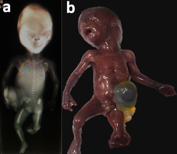
a: Complete body X-ray showing tibial hemimelia present in left extremity. Right lower limb shows the presence of the femur, fibula is not shown due to projection phenomena. Figure 2-b: Fetal specimen, omphalocele, shortening of the left limb and clubfoot can be noted.
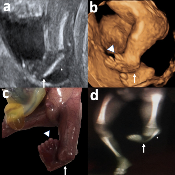
Tibial hemimelia, short fibula (*), clubfoot (arrow), complete pre-axial polydactyly (arrowhead). 3-a: 2D gray scale ultrasound. 3-b: Three-dimensional reconstruction. 3 c: Fetal specimen. 3-d: Postnatal X-ray. Right lower limb displays only tibia because of projection phenomena, fibula is present and of normal characteristics. 2D, two-dimensional.
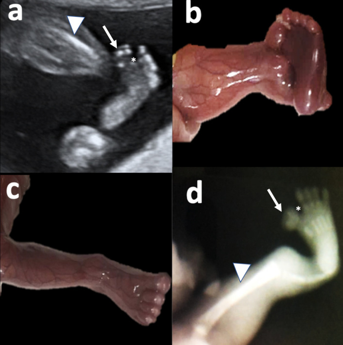
a: Coronal plane of the left foot in 2D Ultrasound. Complete pre-axial polydactyly (arrow), sandal-gap like sign (*), short fibula (arrowhead) and tibial hemimelia are observed. Figure 4 b: Fetal specimen, showing left lower limb with pre-axial polydactyly and sandal-gap sign. Figure 4 c: Fetal specimen, syndactyly in right foot is depicted.Figure 4: X-ray confirming 6 fingers and five metatarsals on the left foot. 2D, two-dimensional.
Similar articles
-
Tibial hemimelia in one of the identical twins.J Pediatr Orthop. 2010 Oct-Nov;30(7):742-5. doi: 10.1097/BPO.0b013e3181edba12. J Pediatr Orthop. 2010. PMID: 20864864
-
Usual Presentation Has Odds: Unilateral Tibial Hemimelia in One of Dizygotic Twins.Cureus. 2021 Jan 21;13(1):e12834. doi: 10.7759/cureus.12834. Cureus. 2021. PMID: 33633877 Free PMC article.
-
[Prevalence of selected congenital anomalies in the Czech Republic: renal and cardiac anomalies and congenital chromosomal aberrations].Epidemiol Mikrobiol Imunol. 2013 Sep;62(3):112-28. Epidemiol Mikrobiol Imunol. 2013. PMID: 24116699 Czech.
-
Systematic radiographic evaluation of tibial hemimelia with orthopedic implications.Pediatr Radiol. 2017 Apr;47(4):473-483. doi: 10.1007/s00247-016-3730-8. Epub 2017 Jan 3. Pediatr Radiol. 2017. PMID: 28050636 Review.
-
Evaluation of prenatally diagnosed structural congenital anomalies.J Obstet Gynaecol Can. 2009 Sep;31(9):875-881. doi: 10.1016/S1701-2163(16)34307-9. J Obstet Gynaecol Can. 2009. PMID: 19941713 Review. English, French.
References
-
- Jones T. Tibial Deficiency. OrthoBullets. 2016. Available from: https://www.orthobullets.com/pediatrics/4058/tibial-deficiency.
Publication types
LinkOut - more resources
Full Text Sources
Other Literature Sources
