. 2021 Jan 5:23:757-768.
doi: 10.1016/j.omtn.2020.12.025. eCollection 2021 Mar 5.

An aptamer interacting with heat shock protein 70 shows therapeutic effects and prognostic ability in serous ovarian cancer

Affiliations

An aptamer interacting with heat shock protein 70 shows therapeutic effects and prognostic ability in serous ovarian cancer

Chang-Ni Lin et al. Mol Ther Nucleic Acids. .

Abstract

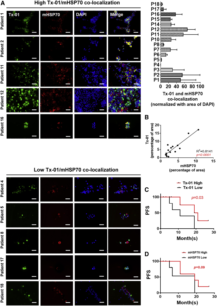
Ovarian cancer (OvCa) is the most lethal gynecologic malignancy owing to its high chemoresistance and late diagnosis, which lead to a poor prognosis. Hence, developing new therapeutic modalities is important for OvCa patient treatment. Our previous results indicated that a novel aptamer, Tx-01, can specifically recognize serous carcinoma cells and tissues. Here, we aim to clarify the clinical role and possible molecular mechanisms of Tx-01 in OvCa. Immunostaining and statistical analysis were performed to detect the interaction of Tx-01 and heat shock protein 70/Notch1 intracellular domain (HSP70/NICD) in OvCa. The in vitro and in vivo experiments were carried out to demonstrate the potential mechanisms of Tx-01. Results show that Tx-01 reduced serous OvCa OVCAR3 cell migration and invasion and inhibited HSP70 nuclear translocation by interrupting the intracellular HSP70/NICD interaction. Furthermore, Tx-01 suppressed serous-type OVCAR3 cell tumor growth in vivo. Tx-01 acts as a prognostic factor through its interaction with membrane-bound HSP70 (mHSP70 that locates on the cell surface without direct interaction to NICD) on ascitic circulating tumor cells (CTCs) and is reported to be involved in natural killer (NK) cell recognition and activation. Our data demonstrated that Tx-01 interacted with HSP70 and showed therapeutic and prognostic effects in serous OvCa. Tx-01 might be a potential inhibitor for use in serous OvCa treatment.

Keywords: Tx-01 aptamer; ascitic CTCs; heat shock protein 70; ovarian cancer; therapeutic and prognostic effects.


Conflict of interest statement

The authors declare no competing interests.

Figures

None
Graphical abstract
Figure 1
Figure 1
Heat shock protein 70 (HSP70) interacted with the Tx-01 aptamer in serous OvCa (A) Immunofluorescence-stained Tx-01 was abundant in the OVCAR3 cells (serous OvCa cell line) compared with that in the IOSE-398 cells (normal ovarian epithelium cells) or TOV-21G cells (clear cell OvCa cell line) staining. (B) 99 candidates interacting with the Tx-01 aptamer were identified by liquid chromatography/tandem mass spectrometry (LC-MS/MS) and included 15 candidates related to ovarian cancer, such as HSP70. (C) HSP70 expression was higher in the serous OvCa cell lines than in the non-serous OvCa cell lines. (D) Tx-01 aptamer-conjugated magnetic beads (Tx-01 aptamer-MBs) and anti-human HSP70 antibody were used to demonstrate the interaction of Tx-01 and HSP70 in OVCAR3 and TOV-21G cells through pull-down assay. The interaction of Tx-01 and HSP70 in the OVCAR3 cells was stronger than that in TOV-21G cells. Random ssDNA-conjugated MBs (random ssDNA-MBs) as the negative control. (E) Confocal microscopy images showed that the colocalization of HSP70 (red) and Tx-01 (green) in the OVCAR3 cells was more obvious than that in the TOV-21G cells. Scale bar, 20 μm. Arrows indicate the colocalization of Tx-01 and HSP70 in the cell membrane. (F) Duolink proximity ligation assay (PLA) showed that the interaction of Tx-01 and HSP70 in the OVCAR3 cells was stronger than that in the TOV-21G cells. (G) The Tx-01 aptamer and HSP70 interaction in the OVCAR3 cells was significantly higher than that in the TOV-21G cells, according to the flow cytometry analyses. ∗∗∗p < 0.01.
Figure 2
Figure 2
Tx-01 is an inhibitor that interrupts the interaction of intracellular HSP70 and the Notch1 intracellular domain (NICD) to suppress OVCAR3 cell migration and invasion (A and B) After Tx-01 (200 nM), DAPT (γ-secretase inhibitor, 5 μg/mL), and KNK437 (HSP70 inhibitor, 100 μM) treatment, cell migration and invasion were significantly reduced in the OVCAR3 cells compared with the untreated cells. Random ssDNA treatment was used as the negative control. Quantification of cell migration (A, right side) and invasion (B, right side) was performed using ImageJ software. ∗p < 0.05; ∗∗p < 0.01; ∗∗∗p < 0.001. (C) Immunofluorescence stain was used to observe the nuclear translocation of HSP70-NICD. After DAPT or KNK437 treatment, HSP70 translocation into the nuclei was obviously reduced. The triangle indicates nuclear HSP70, and the arrow indicates membrane HSP70 in the untreated group (scale bar, 20 μm). Quantification of nuclear HSP70 expression was performed using ImageJ software. ∗p < 0.05; ∗∗p < 0.01; ∗∗∗p < 0.001. (D) Western blot assay was used to confirm that nuclear HSP70 and NICD were obviously reduced after Tx-01, DAPT, or KNK437 treatment. (E) The binding affinity of HSP70 and NICD was significantly reduced after 200 nM, 400 nM, 800 nM, and 1,600 nM Tx-01 treatment. ∗p < 0.05; ∗∗p < 0.01.
Figure 3
Figure 3
Tx-01 inhibited OvCa tumor growth in a xenograft mouse model Serous OvCa patients with high nuclear expression of HSP70 carried poor prognosis. (A and B) Group A, untreated; group B, pretreatment with Tx-01 200 nM for 12 h; group C, Tx-01 200 nM treated twice per week. The OVCAR3 xenograft tumor sizes in groups B and C were significantly smaller and grew more slowly than those of group A (one-way ANOVA; p = 0.0135). The TOV-21G xenograft tumor size and growth rate were not significantly different (one-way ANOVA; p = 0.9713). (C and D) Fluorescence microscopy images showing that nuclear HSP70 was significantly reduced in OVCAR3 xenograft tumors of groups B and C compared with group A OVCAR3 xenograft tumors. The number of apoptotic cells was significantly increased in group C. No significant difference was noted in the TOV-21G xenograft tumors. Scale bar, 100 μm. Quantification of nuclear HSP70 expression was performed using Nuance analysis software based on attachments observed via microscopy. ∗p < 0.05; ∗∗p < 0.01; ∗∗∗p < 0.001. (E) Nuclear HSP70 was observed in serous carcinoma after IHC staining. Images indicate low and high nuclear HSP70. Scale bar, 200 μm. (F) Serous OvCa patients with high nuclear HSP70 expression had poor overall survival (p = 0.0456).
Figure 4
Figure 4
Tx-01 recognized membrane-bound HSP70 (mHSP70) on the circulating tumor cells (CTCs) in ascites and led to favorable prognosis (A) Representative fluorescence microscopy images showing Tx-01 and mHSP70 colocalization in the EpCAM(+) CTCs from serous OvCa patient ascites. Scale bar, 100 μm. Right upper graph shows the summary of Tx-01 and HSP70 colocalization percentage in all 16 ascites samples. (B) Tx-01 expression in the ascitic CTCs was significantly correlated with mHSP70 (p < 0.0001; R2 = 0.8141). (C) High Tx-01 binding in the ascitic CTCs was significantly associated with higher PFS (p = 0.03). (D) High mHSP70 expression in the ascitic CTCs was associated with borderline favorable PFS compared with low mHSP70 expression (p = 0.09). PFS, progression-free survival.
Figure 5
Figure 5
High CD56(+)CD107a(+) NK cells in the ascites led to favorable prognosis for serous OvCa patients (A) After EpCAM-magnetic bead isolation, the EpCAM(–) cells were stained with anti-CD56 and anti-CD107a to detect activated NK cells [CD56(+)CD107a(+)] through flow cytometry. (B) The ascetic CD56(+)CD107a(+) activated NK cells were significantly and positively correlated with CTC mHSP70 expression (R2 = 0.5582; p = 0.0009) and Tx-01 binding (R2 = 0.7221; p < 0.0001). (C) Activated NK cells [CD56(+)CD107a(+)] were significantly associated with PFS (p = 0.04).
Figure 6
Figure 6
Diagram illustrating the dual role of Tx-01 in serous OvCa In the ascitic CTCs, Tx-01 interacts with mHSP70, recognized by ascitic NK cell, and acts as a marker of good prognosis. Intracellular Tx-01 interrupts the interaction of HSP70 and NICD, blocking HSP70 nuclear translocation to reduce cell migration, invasion, and tumor growth.

References

    1. Malvezzi M., Carioli G., Rodriguez T., Negri E., La Vecchia C. Global trends and predictions in ovarian cancer mortality. Ann. Oncol. 2016;27:2017–2025. - PubMed
    1. Siegel R.L., Miller K.D., Jemal A. Cancer statistics, 2019. CA Cancer J. Clin. 2019;69:7–34. - PubMed
    1. Bowtell D.D. The genesis and evolution of high-grade serous ovarian cancer. Nat. Rev. Cancer. 2010;10:803–808. - PubMed
    1. Berchuck A., Secord A.A., Moss H.A., Havrilesky L.J. Maintenance Poly (ADP-ribose) Polymerase Inhibitor Therapy for Ovarian Cancer: Precision Oncology or One Size Fits All? J. Clin. Oncol. 2017;35:3999–4002. - PubMed
    1. Sun H., Zhu X., Lu P.Y., Rosato R.R., Tan W., Zu Y. Oligonucleotide aptamers: new tools for targeted cancer therapy. Mol. Ther. Nucleic Acids. 2014;3:e182. - PMC - PubMed

LinkOut - more resources