. 2021 Jan 16:23:821-834.
doi: 10.1016/j.omtn.2021.01.003. eCollection 2021 Mar 5.

Engineering highly efficient backsplicing and translation of synthetic circRNAs

Affiliations

Engineering highly efficient backsplicing and translation of synthetic circRNAs

Rita M Meganck et al. Mol Ther Nucleic Acids. .

Abstract

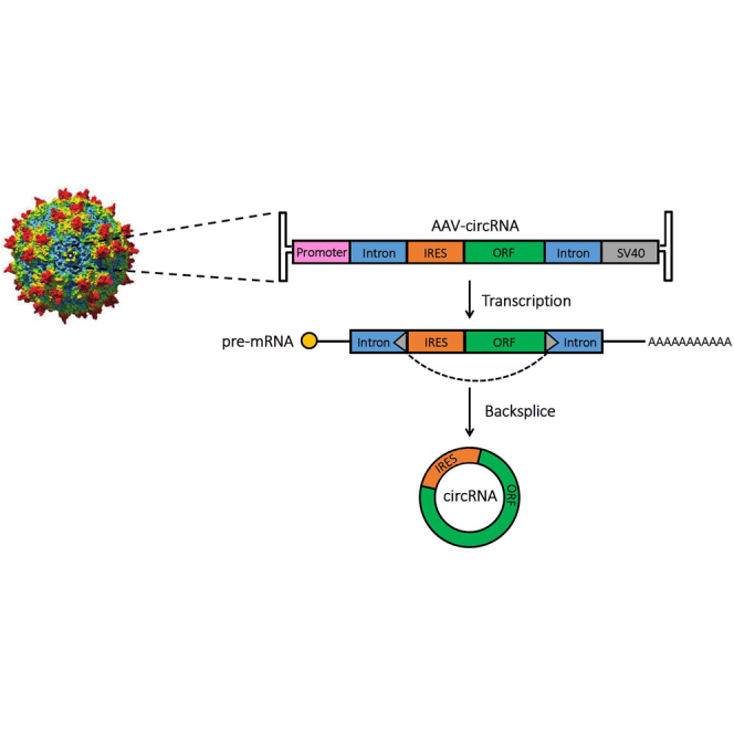
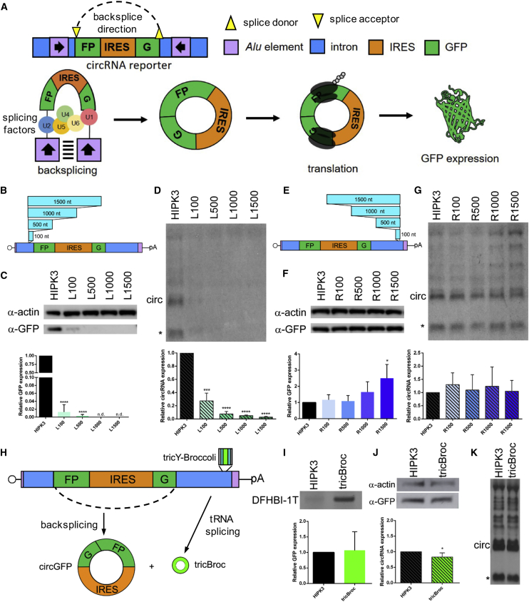
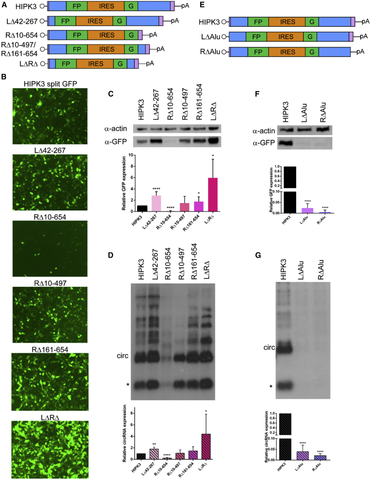
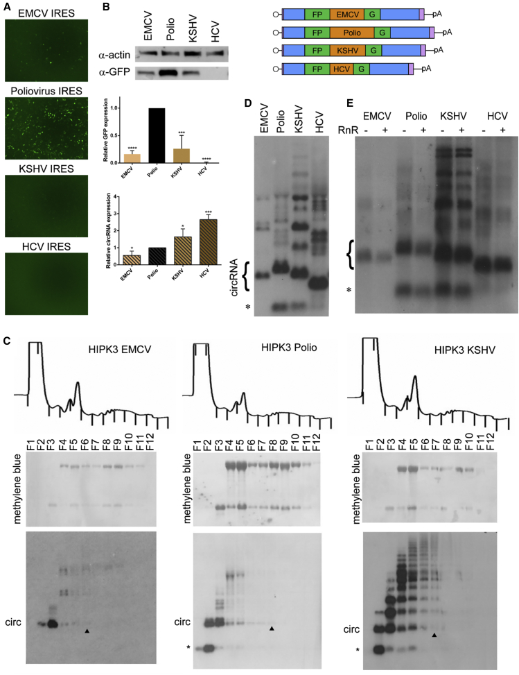
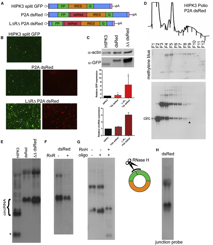
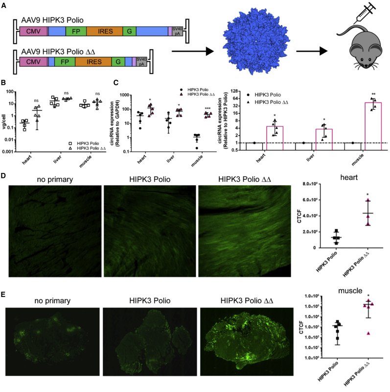
Circular RNAs (circRNAs) are highly stable RNA molecules that are attractive templates for expression of therapeutic proteins and non-coding RNAs. In eukaryotes, circRNAs are primarily generated by the spliceosome through backsplicing. Here, we interrogate different molecular elements including intron type and length, Alu repeats, internal ribosome entry sites (IRESs), and exon length essential for circRNA formation and exploit this information to engineer robust backsplicing and circRNA expression. Specifically, we leverage the finding that the downstream intron can tolerate large inserts without affecting splicing to achieve tandem expression of backspliced circRNAs and tRNA intronic circRNAs from the same template. Further, truncation of selected intronic regions markedly increased circRNA formation in different cell types in vitro as well as AAV-mediated circRNA expression in cardiac and skeletal muscle tissue in vivo. We also observed that different IRES elements and exon length influenced circRNA expression and translation, revealing an exonic contribution to splicing, as evidenced by different RNA species produced. Taken together, these data provide new insight into improving the design and expression of synthetic circRNAs. When combined with AAV capsid and promoter technologies, the backsplicing introns and IRES elements constituting this modular platform significantly expand the gene expression toolkit.


Conflict of interest statement

A.A., W.F.M, and R.M.M. are inventors on patent applications filed on the subject matter of this manuscript. A.A. is a co-founder at TorqueBio, LLC.

Figures

None
Graphical abstract
Figure 1
Figure 1
Distance requirements between Alu element and splice site differ in upstream and downstream introns (A) A reporter was constructed by extracting portions of introns from the HIPK3 gene and placing them around a split GFP reporter exon. Inverted Alu repeats in the introns interact, allowing for backsplicing to occur, forming a circRNA. The presence of an IRES sequence drives translation, leading to GFP protein expression. See Figure S1 for intron sequences. (B) Sequence ranging from 100 nt to 1,500 nt was inserted into the left HIPK3 intron at the indicated position. See also Figure S2. (C and D) Constructs were transfected into HEK293 cells and expression assayed at 4 days post-transfection by (C) western blot analysis, with actin as a loading control (quantification below), and (D) northern blot analysis, probing for GFP sequences (quantification below). (E) Sequence ranging from 100 nt to 1,500 nt was inserted into the right HIPK3 intron at the indicated position. (F and G) Constructs were transfected into HEK293 cells and expression assayed at 4 days post-transfection by (F) western blot analysis, with actin as a loading control (quantification below), and (G) northern blot analysis, probing for GFP sequences (quantification below). (H) Sequences driving formation of a tricRNA containing Broccoli (tricY-Broccoli) were inserted into the same location in the right HIPK3 intron. (I) tricY-Broccoli expression was verified by gel electrophoresis followed by DFHBI-1T staining. (J and K) circRNA formation was assayed by (J) western blot analysis, with actin as a loading control (quantification below left), and (K) northern blot analysis, probing for GFP sequences (quantification left). On northern blots, the asterisk refers to an additional circular band. Western and northern blots were quantified as detailed in Materials and methods and graphed relative to the unchanged HIPK3 intron construct. Student’s t test was performed to test for statistical significance. ∗p < 0.05; ∗∗p < 0.005; ∗∗∗p < 0.0005; ∗∗∗∗p < 0.00005.
Figure 2
Figure 2
Partial truncation of intronic sequences increases circRNA expression (A) Portions of the HIPK3 left and right introns were deleted; deletions are numbered from the start of their respective intron. (B–D) Constructs were transfected into HEK293 cells and expression assayed at 4 days post-transfection by (B) GFP fluorescence, (C) western blot analysis, with actin as a loading control (quantification below), and (D) northern blot analysis, probing for GFP sequences (quantification below). (E) Either the left or right partial Alu element was deleted from the HIPK3 introns. (F and G) Constructs were transfected into HEK293 cells and expression assayed at 4 days post-transfection by (F) western blot analysis, with actin as a loading control, and (G) northern blot analysis, probing for GFP sequences. On northern blots, the asterisk refers to an additional circular band. Western and northern blots were quantified as detailed in Materials and methods and graphed relative to the unchanged HIPK3 intron construct. Student’s t test was performed to test for statistical significance. ∗p < 0.05; ∗∗p < 0.005; ∗∗∗p < 0.0005; ∗∗∗∗p < 0.00005.
Figure 3
Figure 3
IRES elements affect translation efficiency and circRNA expression levels The HIPK3 split GFP construct was created with either the EMCV, Polio, KSHV vFLIP, or HCV IRES elements. (A and B) Constructs were transfected into HEK293 cells and expression assayed at 4 days post-transfection by (A) GFP fluorescence and (B) western blot analysis, with actin as a loading control (quantification below). (C) To assay translation efficiency, the indicated constructs were transfected into HEK293 cells and harvested in cycloheximide followed by a sucrose gradient and fractionation. Top: OD trace of the gradient, with fractions marked by lines. Middle: RNA was extracted from gradients, separated by gel electrophoresis, transferred to a membrane, and stained with methylene blue to visualize the ribosomal RNA. Bottom: the same membrane was probed for GFP sequences. The arrowhead marks the last fraction in which the circRNA is detected. (D) RNA from transfected cells was assayed by northern blot and probed for GFP sequences (quantification left). (E) RNA was digested by RNase R (RnR) followed by northern blotting, probing for GFP sequences. On northern blots, the asterisk refers to an additional circular band. Western and northern blots were quantified as detailed in Materials and methods and graphed relative to the unchanged HIPK3 intron construct. Student’s t test was performed to test for statistical significance. ∗p < 0.05; ∗∗p < 0.005; ∗∗∗p < 0.0005; ∗∗∗∗p < 0.00005.
Figure 4
Figure 4
circRNA size can be increased without loss of expression (A) A self-cleaving P2A peptide followed by the dsRed ORF was added to the exon at the end of the GFP fragment either in the original HIPK3 construct or in the LΔRΔ intron pair. (B and C) Constructs were transfected into HEK293 cells and expression assayed at 4 days post-transfection by (B) GFP and dsRed fluorescence and (C) western blot analysis, with actin as a loading control (quantification below). (D) To assay translation efficiency, the construct was transfected into HEK293 cells and harvested in cycloheximide followed by a sucrose gradient and fractionation. Top: OD trace of the gradient, with fractions marked by lines. Middle: RNA was extracted from gradients, separated by gel electrophoresis, transferred to a membrane, and stained with methylene blue to visualize the ribosomal RNA. Bottom: the same membrane was probed for GFP sequences. The arrowhead marks the last fraction in which the circRNA is detected. (E) RNA from transfected cells was analyzed by northern blot analysis, probing for GFP sequences (quantification above right). (F) RNA was treated with RNase R, then analyzed by northern blot, probing for GFP sequences. (G) RNase H (RnH) digestion was performed with an oligonucleotide targeting the backsplice junction. Samples were analyzed by northern blot and probed against GFP sequences. (H) RNA was analyzed by northern blot and probed with an oligonucleotide spanning the backsplice junction. On northern blots, the asterisk refers to an additional circular band. Western and northern blots were quantified as detailed in Materials and methods and graphed relative to the unchanged HIPK3 intron construct. Student’s t test was performed to test for statistical significance. ∗p < 0.05; ∗∗p < 0.005; ∗∗∗p < 0.0005; ∗∗∗∗p < 0.00005.
Figure 5
Figure 5
The LΔRΔ intron pair afford increased circRNA expression in multiple murine tissues in vivo (A) The indicated constructs were packaged into recombinant AAV9 vectors and injected intravenously into C57BL/6 mice (n = 5) at a dose of 3 × 1011 vector genomes per animal, then harvested 4 weeks post-injection. (B) AAV vector genomes per cell were quantified in each tissue by quantitative PCR (qPCR) for the CMV promoter, normalized to the mouse lamin B2 locus. (C) Quantitative RT-PCR performed with primers amplifying GFP across the backsplice junction revealed increased circRNA expression with the LΔRΔ intron pair. circRNA expression is graphed relative to GAPDH (left) or normalized to HIPK3 Polio expression in each tissue (right). (D) Immunofluorescent staining of sectioned cardiac tissue for GFP expression. The level of GFP expression was quantified by the corrected total cell fluorescence (CTCF) method (right). (E) Immunofluorescent staining of sectioned skeletal muscle tissue showing GFP expression. The level of GFP expression was quantified by the CTCF method (right). Student’s t test was performed to test for statistical significance. ∗p < 0.05; ∗∗p < 0.005; ∗∗∗p < 0.0005; ∗∗∗∗p < 0.00005.

Similar articles

Cited by

References

    1. Jeck W.R., Sorrentino J.A., Wang K., Slevin M.K., Burd C.E., Liu J., Marzluff W.F., Sharpless N.E. Circular RNAs are abundant, conserved, and associated with ALU repeats. RNA. 2013;19:141–157. - PMC - PubMed
    1. Pyle A.M. Group II Intron Self-Splicing. Annu. Rev. Biophys. 2016;45:183–205. - PubMed
    1. Lu Z., Filonov G.S., Noto J.J., Schmidt C.A., Hatkevich T.L., Wen Y., Jaffrey S.R., Matera A.G. Metazoan tRNA introns generate stable circular RNAs in vivo. RNA. 2015;21:1554–1565. - PMC - PubMed
    1. Panda A.C., De S., Grammatikakis I., Munk R., Yang X., Piao Y., Dudekula D.B., Abdelmohsen K., Gorospe M. High-purity circular RNA isolation method (RPAD) reveals vast collection of intronic circRNAs. Nucleic Acids Res. 2017;45:e116. - PMC - PubMed
    1. Zhang Y., Zhang X.O., Chen T., Xiang J.F., Yin Q.F., Xing Y.H., Zhu S., Yang L., Chen L.L. Circular intronic long noncoding RNAs. Mol. Cell. 2013;51:792–806. - PubMed