

Basal Cell Carcinoma With Calcification: Case Report of Calcifying Basal Cell Carcinoma and Review of Calcinosis Cutis Associated With Basal Cell Carcinoma
- PMID: 33614324
- PMCID: PMC7883528
- DOI: 10.7759/cureus.12721
Basal Cell Carcinoma With Calcification: Case Report of Calcifying Basal Cell Carcinoma and Review of Calcinosis Cutis Associated With Basal Cell Carcinoma
Abstract
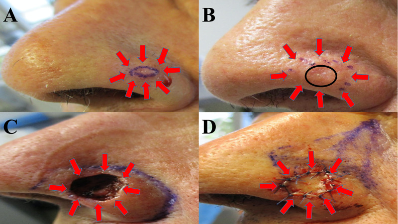
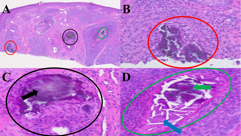
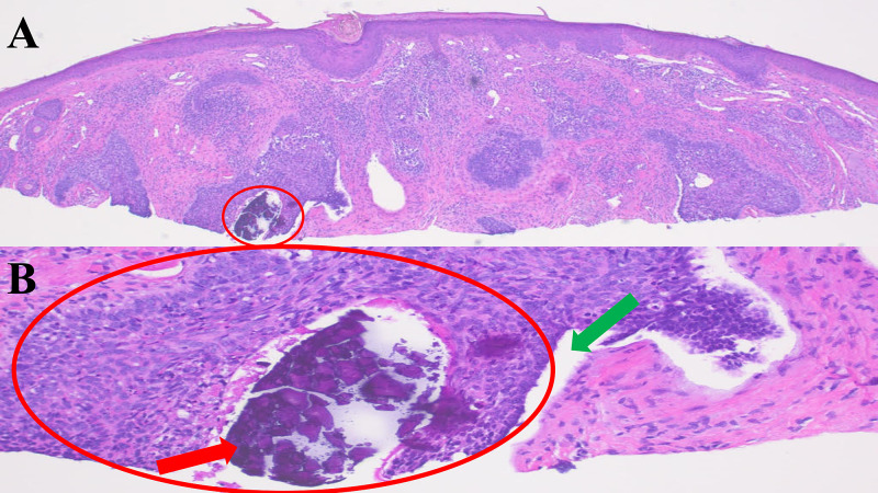
Basal cell carcinoma is the most common cutaneous neoplasm. Calcinosis cutis is the deposition of calcium within the dermis. An 80-year-old man presented with a pearly nodule on his left nasal ala; a shave biopsy confirmed the diagnosis of a nodular basal cell carcinoma with calcinosis cutis, which was removed with Mohs micrographic surgery. The incidence of basal cell carcinoma with calcinosis cutis as well as the classification, identification, and potential origin of calcium deposits in basal cell carcinoma are discussed. Basal cell carcinoma can be associated with calcinosis cutis; indeed, calcifying basal cell carcinoma has a calculated incidence of 14%. There are five categories of calcification in basal cell carcinoma. In addition, calcification observed in cancer-free initial sections of a suspected basal cell carcinoma may be a diagnostic clue that a neoplasm is present in deeper sections of the tissue specimen. Although nodular basal cell carcinoma has the greatest incidence (37%) of calcium deposition, infiltrative (29%) and micronodular (27%) basal cell carcinomas are also frequently associated with calcification; therefore, the presence of calcifying basal cell carcinoma may indicate a more aggressive tumor subtype. Basal cell carcinoma may also be suspected in the differential diagnosis of a superficial breast neoplasm in which calcification is observed in the dermis; in this situation, mammography has been an effective diagnostic approach for identifying the basal cell carcinoma with calcification. The pathogenesis of calcification in basal cell carcinoma remains to be definitively established; however, calcium-binding proteins found in poorly differentiated keratinocytes may contribute to the etiology of basal cell carcinoma with calcification. The treatment of basal cell carcinomas with calcinosis cutis is similar to that of non-calcifying basal cell carcinomas; it is based upon the histologic subtype, the size, and the location of the tumor.
Keywords: basal; calcification; calcinosis; calcium; carcinoma; cell; cutis; histology; mammography; nodular.
Copyright © 2021, Forouzan et al.
Conflict of interest statement
The authors have declared financial relationships, which are detailed in the next section.
Figures
References
-
- Mammographic findings in basal cell carcinoma of the male nipple. Cooper RA, Eilers DB. AJR Am J Roentgenol. 2000;175:1065–1066. - PubMed
-
- Calcifications associated with basal cell carcinoma: prevalence, characteristics, and correlations. Slodkowska EA, Cribier B, Peltre B, Jones DM, Carlson JA. Am J Dermatopathol. 2010;32:557–564. - PubMed
-
- Cutaneous deposition diseases. Part II. Touart DM, Sau P. J Am Acad Dermatol. 1998;39:527–544. - PubMed
-
- Basal cell carcinoma: pathogenesis, epidemiology, clinical features, diagnosis, histopathology, and management. Marzuka AG, Book SE. https://www.ncbi.nlm.nih.gov/pubmed/26029015/ Yale J Biol Med. 2015;88:167–179. - PMC - PubMed
Publication types
LinkOut - more resources
Full Text Sources
Other Literature Sources