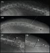
Dural sac localization using myelography and its application to the lumbosacral epidural in dogs
- PMID: 33614431
- PMCID: PMC7830169
- DOI: 10.4314/ovj.v10i4.3
Dural sac localization using myelography and its application to the lumbosacral epidural in dogs
Abstract
Background: The techniques described for the identification of the lumbosacral (LS) epidural space in dogs do not guarantee the needle position or an accidental subarachnoid puncture, especially in small size dogs.
Aim: To determine the relationship between body weight and the location of the dural sac (DS) using myelography in dogs, and to determine the possibility of subarachnoid puncture during LS epidural based on the position of the DS.
Methods: Four masked observers evaluated 70 myelographic studies of dogs, annotating the vertebrae where the DS ended, if it was localized before or after the LS space, and if accidental subarachnoid puncture during LS epidural injection was possible (yes/no). Body weight (kg) was categorized into: less than 10 kg, between 10 and 20 kg, and more than 20 kg and was also converted to body surface area (BSA) as a continuous variable.
Results: The DS ended at the LS space or caudally in 50% of dogs. There was a statistically significant difference between the position of the DS and the dog's BSA (p = 0.001). The DS ended caudal to the LS space in 72.7% of dogs weighing <10 kg, in 25% of dogs between 10 and 20 kg and in 15% of dogs in the >20 kg category. The observers considered a possible subarachnoid puncture during LS epidural in 69.7% of patients <10 kg, 16.6% on those between 10 and 20 kg, and in 11.7% of the dogs >20 kg.
Conclusion: The DS ended caudal to the LS space in almost 3/4 dogs in the <10 kg category, so accidental subarachnoid puncture during LS epidural is highly possible in this weight range.
Keywords: Body weight; Dog; Dural sac; Lumbosacral epidural; Myelography.
Conflict of interest statement
The authors declare that there is no conflict of interest.
Figures

References
-
- Adami C, Gendron K. What is the evidence? The issue of verifying correct needle position during epidural anaesthesia in dogs. Vet. Anaesth. Analg. 2017;44:212–218. - PubMed
-
- Campoy L, Read M, , Peralta S. Canine and feline local anesthetic and analgesic techniques. In: Thurmon J.C, William J.T, Grimm K.A, Lumb V.L, Jones E.W, editors. Lumb and Jones Veterinary Anesthesia and Analgesia. 5th. Iowa, IA: Wiley-Blackwell; 2015. pp. 847–851.
-
- Concetto S.D, Mandsager R.E, Riebold T.W, Stiger-Vanegas S.M, Killos M. Effect of hind limb position on the craniocaudal length of the lumbosacral space in anesthetized dogs. Vet. Anaesth. Analg. 2012;39:99–105. - PubMed
-
- Duke -Novakovski T. Pain management II: local and regional anaesthetic techniques. In: Duke Novakovski T, Vries M, Seymour C, editors. BSAVA manual of canine and feline anaesthesia and analgesia. 3rd. Gloucester, UK: Small Animal Veterinary Association; 2016. pp. 152–155.
-
- Fletcher T.F. Spinal cord and meninges. In: Evans H.E, Miller M.E, De Lahunta A, editors. Miller’s anatomy of the dog. 4th. St. Louis, MO: Elsevier Saunders; 2013. pp. 59–610.
MeSH terms
LinkOut - more resources
Full Text Sources
Other Literature Sources
