

A pilot study on vitrectomy combined with scleral shortening for eyes with myopic macular retinoschisis-2y follow-up results
- PMID: 33614456
- PMCID: PMC7840361
- DOI: 10.18240/ijo.2021.02.13
A pilot study on vitrectomy combined with scleral shortening for eyes with myopic macular retinoschisis-2y follow-up results
Abstract
Aim: To evaluate the effect of vitrectomy combined with scleral shortening for eyes with myopic macular retinoschisis.
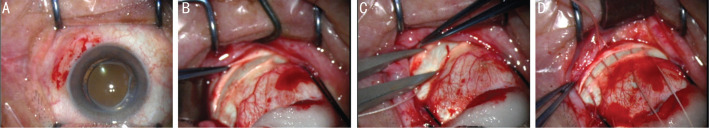
Methods: Thirty-seven patients with myopic macular retinoschisis who underwent pars plana vitrectomy (PPV) combined with scleral shortening were reviewed. Axial length (AL), the height of macular retinoschisis, the height of retinal detachment if existed, the diameter of macular hole if existed and best corrected visual acuity (BCVA) were obtained. The preoperative and postoperative parameters were compared.
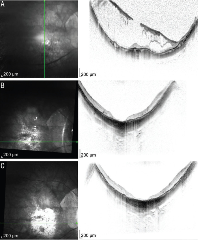
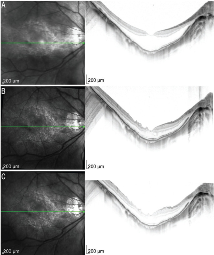
Results: At postoperative 24mo, the mean AL and height of macular retinoschisis were reduced significantly by 0.79 mm and 256.51 µm (t=8.064, P<0.0001; Z=-5.086, P<0.0001) respectively. In addition, the mean height of retinal detachment and diameter of macular hole were also reduced significantly by 365.38 µm and 183.68 µm (Z=-4.457, P=0.000008; Z=-2.983, P=0.003) respectively. Meanwhile, the postoperative BCVA was improved markedly (Z=-2.126, P=0.033).
Conclusion: Vitrectomy combined with scleral shortening is an effective surgical method for eyes with myopic macular retinoschisis, whether or not macular hole and retinal detachment are present.
Keywords: macula; myopia; retinoschisis; scleral shortening; vitrectomy.
International Journal of Ophthalmology Press.
Figures
Similar articles
-
Posterior scleral reinforcement and vitrectomy for myopic foveoschisis in extreme myopia.Retina. 2015 Feb;35(2):351-7. doi: 10.1097/IAE.0000000000000313. Retina. 2015. PMID: 25111687
-
Retinoschisis: A Predictive Factor in Vitrectomy for Lamellar Macular Holes in Highly Myopic Eyes.Ophthalmologica. 2019;242(4):208-213. doi: 10.1159/000500927. Epub 2019 Jul 17. Ophthalmologica. 2019. PMID: 31315117
-
Pars plana vitrectomy in patients with myopic macular retinoschisis.Br J Ophthalmol. 2014 Apr;98(4):534-7. doi: 10.1136/bjophthalmol-2013-304578. Epub 2014 Jan 10. Br J Ophthalmol. 2014. PMID: 24414402
-
Macular buckle technique in myopic traction maculopathy: a 16-year review of the literature and a comparison with vitreous surgery.Graefes Arch Clin Exp Ophthalmol. 2018 May;256(5):863-877. doi: 10.1007/s00417-018-3947-3. Epub 2018 Mar 28. Graefes Arch Clin Exp Ophthalmol. 2018. PMID: 29589106 Review.
-
The effectiveness and safety of posterior scleral reinforcement with vitrectomy for myopic foveoschisis treatment: a systematic review and meta-analysis.Graefes Arch Clin Exp Ophthalmol. 2020 Feb;258(2):257-271. doi: 10.1007/s00417-019-04550-5. Epub 2019 Dec 10. Graefes Arch Clin Exp Ophthalmol. 2020. PMID: 31823060
Cited by
-
Visualized analysis of research on myopic traction maculopathy based on CiteSpace.Int J Ophthalmol. 2023 Dec 18;16(12):2117-2124. doi: 10.18240/ijo.2023.12.26. eCollection 2023. Int J Ophthalmol. 2023. PMID: 38111942 Free PMC article.
References
-
- Shimada N, Ohno-Matsui K, Baba T, Futagami S, Tokoro T, Mochizuki M. Natural course of macular retinoschisis in highly myopic eyes without macular hole or retinal detachment. Am J Ophthalmol. 2006;142(3):497–500. - PubMed
-
- Shimada N, Tanaka Y, Tokoro T, Ohno-Matsui K. Natural course of myopic traction maculopathy and factors associated with progression or resolution. Am J Ophthalmol. 2013;156(5):948–957.e1. - PubMed
-
- Wu PC, Chen YJ, Chen YH, Chen CH, Shin SJ, Tsai CL, Kuo HK. Factors associated with foveoschisis and foveal detachment without macular hole in high myopia. Eye (Lond) 2009;23(2):356–361. - PubMed
-
- Ikuno Y, Gomi F, Tano Y. Potent retinal arteriolar traction as a possible cause of myopic foveoschisis. Am J Ophthalmol. 2005;139(3):462–467. - PubMed
LinkOut - more resources
Full Text Sources
Other Literature Sources
Miscellaneous