. 2021 Feb 3:10:617310.
doi: 10.3389/fcimb.2020.617310. eCollection 2020.

A New Family of Bacteriolytic Proteins in Dictyostelium discoideum

Affiliations

A New Family of Bacteriolytic Proteins in Dictyostelium discoideum

Cyril Guilhen et al. Front Cell Infect Microbiol. .

Abstract

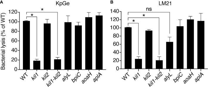
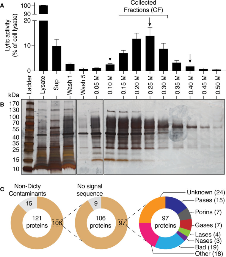
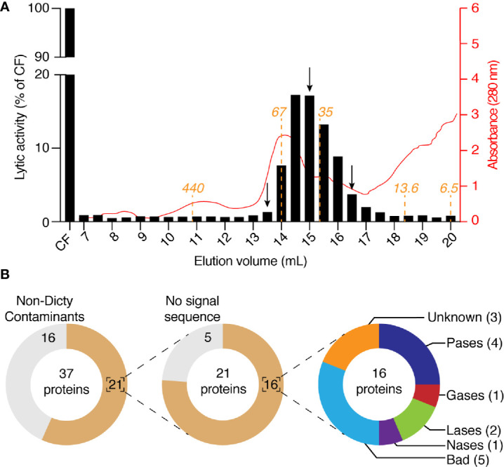
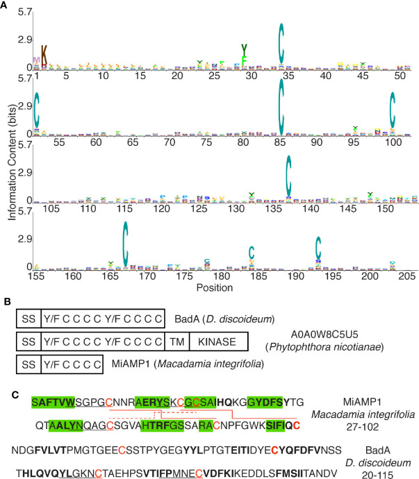
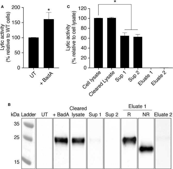
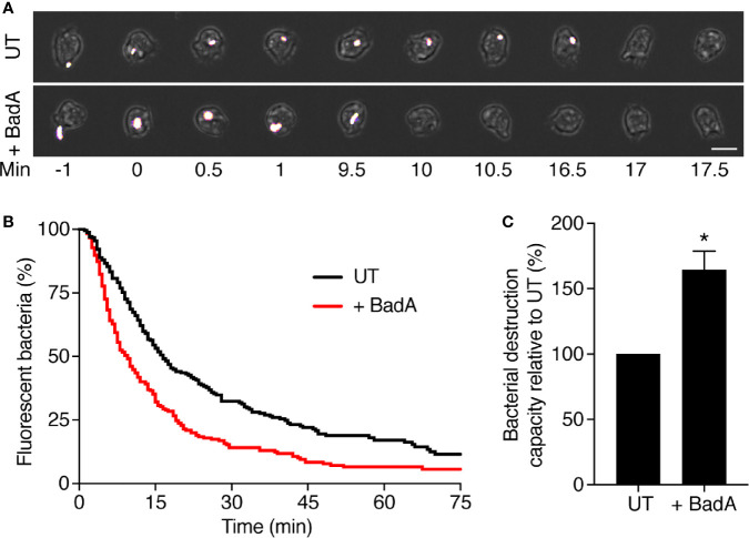
Phagocytic cells ingest and destroy bacteria efficiently and in doing so ensure the defense of the human body against infections. Phagocytic Dictyostelium discoideum amoebae represent a powerful model system to study the intracellular mechanisms ensuring destruction of ingested bacteria in phagosomes. Here, we discovered the presence of a bacteriolytic activity against Klebsiella pneumoniae in cellular extracts from D. discoideum. The bacteriolytic activity was detected only at a very acidic pH mimicking the conditions found in D. discoideum phagosomes. It was also strongly decreased in extracts of kil1 KO cells that were previously described to kill inefficiently internalized bacteria, suggesting that the activity observed in vitro is involved in killing of bacteria in phagosomes. We purified a fraction enriched in bacteriolytic activity where only 16 proteins were detected and focused on four proteins selectively enriched in this fraction. Three of them belong to a poorly characterized family of D. discoideum proteins exhibiting a DUF3430 domain of unknown function and were named BadA (Bacteriolytic D. discoideum A), BadB, and BadC. We overexpressed the BadA protein in cells, and the bacteriolytic activity increased concomitantly in cell extracts. Conversely, depletion of BadA from cell extracts decreased significantly their bacteriolytic activity. Finally, in cells overexpressing BadA, bacterial killing was faster than in parental cells. Together these results identify BadA as a D. discoideum protein required for cellular bactericidal activity. They also define a new strategy to identify and characterize bactericidal proteins in D. discoideum cells.

Keywords: Bacteriolytic D. discoideum A (BadA); Dictyostelium discoideum; Klebsiella pneumoniae; bacteriolytic proteins; intracellular killing; protein purification.


Conflict of interest statement

The authors declare that the research was conducted in the absence of any commercial or financial relationships that could be construed as a potential conflict of interest.

Figures

Figure 1
Figure 1
Bacteriolytic activities in D. discoideum cell extracts. (A) A cell extract from WT D. discoideum was serially diluted (from 1/8 to 1/256) in lysis buffer and then mixed with K. pneumoniae KpGe bacteria at pH2. The bacteriolytic activity was monitored over time by spectrophotometry at 450 nm (OD450). Results are expressed as a percentage of OD450 at time 0. D. discoideum cell extracts lysed K. pneumoniae KpGe bacteria at pH2. This is a set of data from a representative experiment. (B, C) The bacteriolytic activity was measured as described in (A) over a range of pH from 1 to 7. The OD450 values after 120 min of incubation were used to determine the effect of pH on the bacteriolytic activity of the cell extract (dilution 1/32). The bacteriolytic activity against K. pneumoniae KpGe (B) and LM21 (C) bacteria was maximal at pH1.5. No activity was observed above 2.5. Approximatively 25% of bacteria spontaneously lysed at pH1 and 1.5 (mean ± SEM; N = 3 independent experiments; *: p <.05; student t test). (D) Bacteria were washed once in phosphate buffer at the indicated pH and 10 μl were spotted on a LB agar plate to measure bacterial viability. Both K. pneumoniae KpGe and LM21 bacteria were dead at acidic pH (1.5 or 2) before the beginning of the bacterial lysis assay. Thus, in the conditions where the bacterial lysis is assessed, bacteria are dead.
Figure 2
Figure 2
The D. discoideum bacteriolytic activity is sensitive to high temperature. D. discoideum cell extracts (dilution: 1/8) were incubated for 1 h at the indicated temperature. They were then mixed with K. pneumoniae KpGe bacteria at pH2 and the bacteriolytic activity was monitored overtime by spectrophotometry at 450 nm. Results are expressed as a percentage of OD450 at time 0. This is a set of data from a representative experiment.
Figure 3
Figure 3
The D. discoideum bacteriolytic activity is more efficient against bacteria with disrupted cell walls. D. discoideum cell extracts (dilution: 1/32) were mixed with either WT or KO waaQ K. pneumoniae KpGe bacteria at pH2 and the bacteriolytic activity was monitored overtime by spectrophotometry at 450 nm. Results are expressed as a percentage of OD450 at time 0 (mean ± SEM; N = 4 independent experiments; *: p <.05; student t test).
Figure 4
Figure 4
The bacteriolytic activity of kil1 KO D. discoideum cells is weaker than in WT cells. Cell extracts (diluted either 8, 16, 32, 64, 128, or 256 times) from D. discoideum WT, kil1, kil2, kil1-kil2, alyL, bpiC, aoaH, or aplA KO strains were mixed with either K. pneumoniae KpGe (A) or K. pneumoniae LM21 (B) bacterial strains and bacterial lysis assessed. Results are expressed as a percentage of OD450 values obtained with WT amoeba after 2 h of incubation (mean ± SEM; *p < .01; student t test; for panel A: kil1: N = 14, kil2: N = 11, kil1-kil2: N = 12, alyL and bpiC: N = 6, aoaH: N = 7, aplA: N = 5; for panel B: N = 4).
Figure 5
Figure 5
Purification by anionic exchange fractionation of bacteriolytic proteins. WT D. discoideum cell lysate was mixed with an anionic exchange resin at pH3. The anionic resin was washed five times and proteins fixed to the resin were eluted with a gradient of NaCl from 0.05 M to 0.5 M of NaCl. Unattached proteins were recovered in the supernatant (sup). (A) each fraction was tested for its bacteriolytic activity against K. pneumoniae KpGe bacteria. Results are expressed as a percentage of OD450 values obtained with total cell lysate after 2 h of incubation (mean ± SEM; N = 7 independent experiments). (B) Silver nitrate stained SDS PAGE gel showing protein composition pattern of each of the obtained fractions. 0.75 μl over the 1,700 μl of either the total cell lysate or supernatant were loaded in the gel. Fifteen μl over the 1,000 μl of washing steps or the 200 μl of each of the elution were loaded. Molecular mass markers are indicated at the left. (C) Protein composition of three fractions (indicated by a black arrow; e.g., 0.10, 0.25, and 0.40 M) were analyzed by mass spectrometry. Among the 121 proteins identified in the three samples, 15 were non- D. discoideum proteins (e.g., human keratin) and nine do not display a signal sequence. Of the 97 remaining D. discoideum proteins, 15 were proteases (Pases), 7 were porins, 7 were glycosidases (Gases), 4 were lipases (Lases), and 3 were nucleases (Nases). Finally, 43 had an unknown function (including 19 Bad proteins), and 18 had non-lytic functions.
Figure 6
Figure 6
Purification by size-exclusion chromatography of bacteriolytic proteins. (A) The four fractions with the highest activity in Figure 5A (e.g., 0.15, 0.20, 0.25, and 0.30) were collected, mixed, buffered at pH7 and loaded on a size-exclusion chromatographic column. The column (Superdex 200; 10 x 300 mm) was equilibrated with NaP buffer at pH7 and eluted with the same buffer at a rate of 18 ml/h and 0.5 ml by fraction. Each of the obtained fractions was then tested for their bacteriolytic activity against K. pneumoniae KpGe bacteria. Results are from a single experiment, and are expressed as a percentage of OD450 values obtained with the four collected fractions after 2 h of incubation. Relative protein abundance in each of the obtained fractions was monitored by spectrophotometry at 280 nm. The predicted molecular weight of proteins eluted from the Superdex 200 column were indicated in orange. (B) Protein composition of three fractions (indicated by a black arrow; e.g., 13.5, 15, and 16.5 ml) were analyzed by mass spectrometry. Among the 37 proteins identified in the three samples, 16 were non- D. discoideum proteins and 5 do not display a signal sequence. Of the 16 remaining D. discoideum proteins, 4 were proteases (Pases), 1 was glycosidase (Gases), 2 were lipases (Lases), and 1 was nuclease (Nases). Finally, eight had an unknown function (including five Bad proteins).
Figure 7
Figure 7
The DUF3430 domain. (A) Positions 1 to 205 of the DUF3430 profile HMM from Pfam (PF11912, Pfam 33.1), produced using Skylign (Wheeler et al., 2014) and showing the conserved residues at specific positions. The profile HMM was constructed using hmmbuild (default parameters) from HMMER 3.1 on the Pfam full alignment (163 sequences). (B) Schematic organization of domains with conserved aromatics and cysteine residues in BadA, a member of a large family of Phytophthora proteins and the plant MiAMP1 protein. (C) Sequence and structure of the MiAMP1 protein and the first half of the DUF3430 motif of BadA. Residues shown to be engaged in β-strands are highlighted in green, residues predicted by SOPMA to be engaged in β-strands are in bold, Y/W/FxxxxC motifs are underlined, disulfide bonds are indicated with red lines.
Figure 8
Figure 8
The protein BadA is involved in the lysis of K. pneumoniae KpGe bacteria. (A) Total cell lysate from untransfected (UT-cells) and BadA-ALFA transfected D. discoideum cell were mixed with Kp Ge bacteria at pH2 and the bacteriolytic activity was monitored overtime by spectrophotometry at 450 nm. Results are expressed as a percentage of OD450 values obtained with untransfected cell lysate after 2 h of incubation (mean ± SEM; N = 10 independent experiments). Cell lysate from BadA-ALFA transfected D. discoideum cell was incubated with uncoupled protein A agarose beads. The resulting supernatant (cleared lysate) was subjected to 2 successive immunoprecipitations with the ALFA selector PE resin (Götzke et al., 2019) to obtain respectively supernatants (sup) 1 and 2. Proteins attached to the resin were finally eluted by competition using the ALFA peptide. (B) UT and BadA-ALFA transfected cell lysates, as well as the cleared lysate, sup 1, sup 2 and eluates 1 and 2 fractions from IP were separated on an SDS-polyacrylamide gel in reducing (R) or non-reducing (NR) conditions, transferred to a nitrocellulose membrane and revealed using an anti-ALFA antibody. (C) BadA-ALFA transfected cell lysate, as well as the cleared lysate, sup 1, sup 2, eluates 1 and 2 fractions from IP were tested for their bacteriolytic activity against K. pneumoniae KpGe bacteria. Results are expressed as a percentage of OD450 values obtained with transfected cell lysate after 2 h of incubation (mean ± SEM; N = 5 independent experiments; *p < .01; student t test).
Figure 9
Figure 9
BadA is involved in the lysis of K. pneumoniae KpGe bacteria in phagosomes. To visualize ingestion and intracellular destruction of individual bacterial, D. discoideum cells were incubated with GFP-expressing K. pneumoniae at a ratio of 1:3 for 2 h. Cells were imaged every 30 s by phase contrast and fluorescence microscopy. (A) Successive images of untransfected D. discoideum cells ingesting (t = 0) and destroying (t = 17 min) individual K. pneumoniae bacterium. Below, a BadA-ALFA transfected cell destroyed a K. pneumoniae bacterium 10 min after ingestion. Scale bar: 10 μm. (B) The time between ingestion and fluorescence extinction was determined for each bacterium and the probability of remaining fluorescent was represented as a function of time after ingestion. Bacterial destruction was analyzed in UT and BadA-ALFA transfected cells. The curves shown were obtained by pooling the results of eight independent experiments. (C) Quantification of bacterial destruction capacity of BadA-ALFA transfected cells relative to UT cells. In each independent experiment, a UT control was included, and the bacterial destruction capacity of BadA-ALFA cells was determined using the following formula: (AUCUT/AUC+BadA)x100; (mean ± SEM; N = 8 independent experiments; total number of events observed: UT = 240, + BadA = 238; *p < .005; student t test).

Similar articles

Cited by

References

    1. Andrä J., Herbst R., Leippe M. (2003). Amoebapores, archaic effector peptides of protozoan origin, are discharged into phagosomes and kill bacteria by permeabilizing their membranes. Dev. Comp. Immunol. 27, 291–304. 10.1016/S0145-305X(02)00106-4 - DOI - PubMed
    1. Ashburner M., Ball C. A., Blake J. A., Botstein D., Butler H., Cherry J. M., et al. (2000). Gene Ontology: tool for the unification of biology. Nat. Genet. 25, 25–29. 10.1038/75556 - DOI - PMC - PubMed
    1. Balestrino D., Ghigo J.-M., Charbonnel N., Haagensen J. A. J., Forestier C. (2008). The characterization of functions involved in the establishment and maturation of Klebsiella pneumoniae in vitro biofilm reveals dual roles for surface exopolysaccharides. Environ. Microbiol. 10, 685–701. 10.1111/j.1462-2920.2007.01491.x - DOI - PubMed
    1. Benghezal M., Fauvarque M.-O., Tournebize R., Froquet R., Marchetti A., Bergeret E., et al. (2006). Specific host genes required for the killing of Klebsiella bacteria by phagocytes. Cell. Microbiol. 8, 139–148. 10.1111/j.1462-5822.2005.00607.x - DOI - PubMed
    1. Bodinier R., Leiba J., Sabra A., Jauslin T. N., Lamrabet O., Guilhen C., et al. (2020). LrrkA, a kinase with leucine-rich repeats, links folate sensing with Kil2 activity and intracellular killing. Cell. Microbiol. 22, e13129. 10.1111/cmi.13129 - DOI - PMC - PubMed

Publication types

Substances