

Recent Advances in Our Understanding of the Diversity and Roles of Chaperone-Usher Fimbriae in Facilitating Salmonella Host and Tissue Tropism
- PMID: 33614531
- PMCID: PMC7886704
- DOI: 10.3389/fcimb.2020.628043
Recent Advances in Our Understanding of the Diversity and Roles of Chaperone-Usher Fimbriae in Facilitating Salmonella Host and Tissue Tropism
Abstract
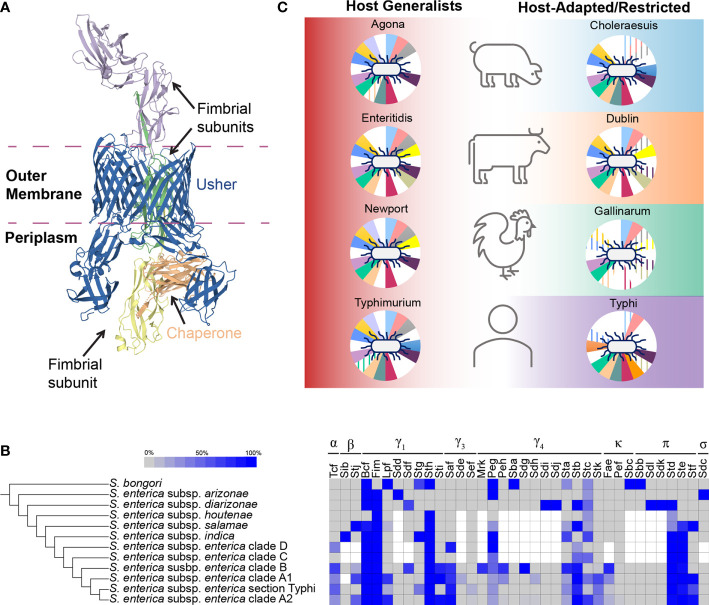
Salmonella enterica is one of the most diverse and successful pathogens, representing a species with >2,600 serovars with a variety of adaptations that enable colonization and infection of a wide range of hosts. Fimbriae, thin hair-like projections that cover the surface of Salmonella, are thought to be the primary organelles that mediate Salmonella's interaction with, and adherence to, the host intestinal epithelium, representing an important step in the infection process. The recent expansion in genome sequencing efforts has enabled the discovery of novel fimbriae, thereby providing new perspectives on fimbrial diversity and distribution among a broad number of serovars. In this review, we provide an updated overview of the evolutionary events that shaped the Salmonella chaperone-usher fimbriome in light of recent phylogenetic studies describing the population structure of Salmonella enterica. Furthermore, we discuss the complexities of the chaperone-usher fimbriae-mediated host-pathogen interactions and the apparent redundant roles of chaperone-usher fimbriae in host and tissue tropism.
Keywords: Salmonella; adhesin Salmonella; chaperone-usher; fimbriae; host-pathogen interaction.
Copyright © 2021 Cheng and Wiedmann.
Conflict of interest statement
The authors declare that the research was conducted in the absence of any commercial or financial relationships that could be construed as a potential conflict of interest.
Figures
Similar articles
-
The Number and Type of Chaperone-Usher Fimbriae Reflect Phylogenetic Clade Rather than Host Range in Salmonella.mSystems. 2022 Jun 28;7(3):e0011522. doi: 10.1128/msystems.00115-22. Epub 2022 Apr 25. mSystems. 2022. PMID: 35467401 Free PMC article. Review.
-
Functional Analysis of the Chaperone-Usher Fimbrial Gene Clusters of Salmonella enterica serovar Typhi.Front Cell Infect Microbiol. 2018 Feb 8;8:26. doi: 10.3389/fcimb.2018.00026. eCollection 2018. Front Cell Infect Microbiol. 2018. PMID: 29473020 Free PMC article.
-
Type 1 fimbria and P pili: regulatory mechanisms of the prototypical members of the chaperone-usher fimbrial family.Arch Microbiol. 2024 Aug 10;206(9):373. doi: 10.1007/s00203-024-04092-3. Arch Microbiol. 2024. PMID: 39127787 Free PMC article. Review.
-
Chaperone-usher fimbriae in a diverse selection of Gallibacterium genomes.BMC Genomics. 2014 Dec 12;15(1):1093. doi: 10.1186/1471-2164-15-1093. BMC Genomics. 2014. PMID: 25495603 Free PMC article.
-
Diversification of the Salmonella fimbriae: a model of macro- and microevolution.PLoS One. 2012;7(6):e38596. doi: 10.1371/journal.pone.0038596. Epub 2012 Jun 12. PLoS One. 2012. PMID: 22701679 Free PMC article.
Cited by
-
Comprehensive blueprint of Salmonella genomic plasticity identifies hotspots for pathogenicity genes.PLoS Biol. 2024 Aug 7;22(8):e3002746. doi: 10.1371/journal.pbio.3002746. eCollection 2024 Aug. PLoS Biol. 2024. PMID: 39110680 Free PMC article.
-
Differences between the global transcriptomes of Salmonella enterica serovars Dublin and Cerro infecting bovine epithelial cells.BMC Genomics. 2022 Jul 8;23(1):498. doi: 10.1186/s12864-022-08725-z. BMC Genomics. 2022. PMID: 35804292 Free PMC article.
-
Genomic and phenotypic characterization of Salmonella enterica serovar Kentucky.Microb Genom. 2023 Sep;9(9):001089. doi: 10.1099/mgen.0.001089. Microb Genom. 2023. PMID: 37750759 Free PMC article.
-
Comparative genome analysis of Salmonella enterica serovar Gallinarum biovars Pullorum and Gallinarum decodes strain specific genes.PLoS One. 2021 Aug 19;16(8):e0255612. doi: 10.1371/journal.pone.0255612. eCollection 2021. PLoS One. 2021. PMID: 34411120 Free PMC article.
-
Antimicrobial Resistance and Genomic Characterization of Salmonella Serovars Typhimurium and 4,[5],12:i:- in Huzhou, China.Infect Drug Resist. 2025 May 31;18:2765-2777. doi: 10.2147/IDR.S521802. eCollection 2025. Infect Drug Resist. 2025. PMID: 40469478 Free PMC article.
References
-
- Aviv G., Elpers L., Mikhlin S., Cohen H., Vitman Zilber S., Grassl G. A., et al. (2017). The plasmid-encoded Ipf and Klf fimbriae display different expression and varying roles in the virulence of Salmonella enterica serovar Infantis in mouse vs. avian hosts. PLoS Path. 13, e1006559–e1006559. 10.1371/journal.ppat.1006559 - DOI - PMC - PubMed
-
- Azriel S., Goren A., Shomer I., Aviv G., Rahav G., Gal-Mor O. (2017). The Typhi colonization factor (Tcf) is encoded by multiple non-typhoidal Salmonella serovars but exhibits a varying expression profile and interchanging contribution to intestinal colonization. Virulence 8, 1791–1807. 10.1080/21505594.2017.1380766 - DOI - PMC - PubMed
-
- Bäumler A. J., Tsolis R. M., Bowe F. A., Kusters J. G., Hoffmann S., Heffron F. (1996. a). The pef fimbrial operon of Salmonella Typhimurium mediates adhesion to murine small intestine and is necessary for fluid accumulation in the infant mouse. Infect. Immun. 64, 61–68. 10.1128/IAI.64.1.61-68.1996 - DOI - PMC - PubMed
Publication types
MeSH terms
Substances
LinkOut - more resources
Full Text Sources
Other Literature Sources