

Graphene and its Derivatives-Based Optical Sensors
- PMID: 33614600
- PMCID: PMC7892452
- DOI: 10.3389/fchem.2021.615164
Graphene and its Derivatives-Based Optical Sensors
Abstract
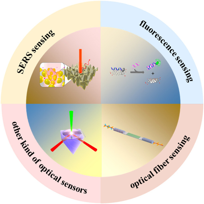
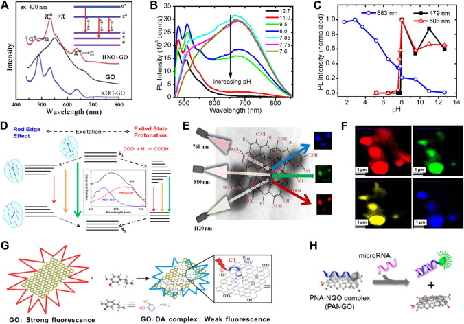
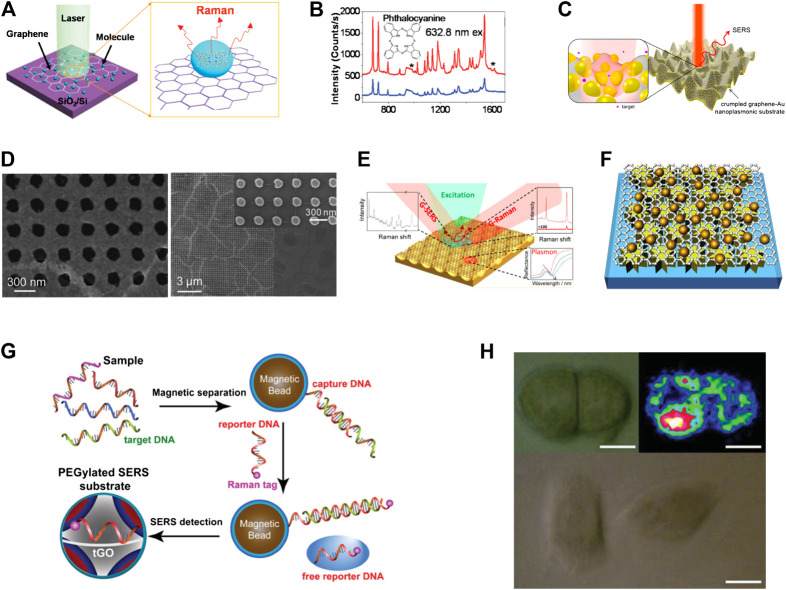
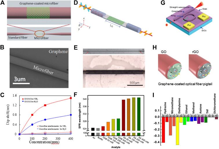
Being the first successfully prepared two-dimensional material, graphene has attracted extensive attention from researchers due to its excellent properties and extremely wide range of applications. In particular, graphene and its derivatives have displayed several ideal properties, including broadband light absorption, ability to quench fluorescence, excellent biocompatibility, and strong polarization-dependent effects, thus emerging as one of the most popular platforms for optical sensors. Graphene and its derivatives-based optical sensors have numerous advantages, such as high sensitivity, low-cost, fast response time, and small dimensions. In this review, recent developments in graphene and its derivatives-based optical sensors are summarized, covering aspects related to fluorescence, graphene-based substrates for surface-enhanced Raman scattering (SERS), optical fiber biological sensors, and other kinds of graphene-based optical sensors. Various sensing applications, such as single-cell detection, cancer diagnosis, protein, and DNA sensing, are introduced and discussed systematically. Finally, a summary and roadmap of current and future trends are presented in order to provide a prospect for the development of graphene and its derivatives-based optical sensors.
Keywords: fluorescence; graphene; optical fiber; optical sensors; surface-enhanced Raman scattering.
Copyright © 2021 Gao, Cheng, Jiang, Li and Xing.
Conflict of interest statement
The authors declare that the research was conducted in the absence of any commercial or financial relationships that could be construed as a potential conflict of interest.
Figures
Similar articles
-
Optical Biosensor Based on Graphene and Its Derivatives for Detecting Biomolecules.Int J Mol Sci. 2022 Sep 16;23(18):10838. doi: 10.3390/ijms231810838. Int J Mol Sci. 2022. PMID: 36142748 Free PMC article. Review.
-
Recent progress on graphene-based substrates for surface-enhanced Raman scattering applications.J Mater Chem B. 2018 Jun 28;6(24):4008-4028. doi: 10.1039/c8tb00902c. Epub 2018 May 29. J Mater Chem B. 2018. PMID: 32255147
-
Review of Polarization Optical Devices Based on Graphene Materials.Int J Mol Sci. 2020 Feb 26;21(5):1608. doi: 10.3390/ijms21051608. Int J Mol Sci. 2020. PMID: 32111096 Free PMC article. Review.
-
Applications of graphene and its derivatives in intracellular biosensing and bioimaging.Analyst. 2016 Aug 7;141(15):4541-53. doi: 10.1039/c6an01090c. Epub 2016 Jul 4. Analyst. 2016. PMID: 27373227 Review.
-
Raman and Fluorescence Enhancement Approaches in Graphene-Based Platforms for Optical Sensing and Imaging.Nanomaterials (Basel). 2021 Mar 5;11(3):644. doi: 10.3390/nano11030644. Nanomaterials (Basel). 2021. PMID: 33808013 Free PMC article. Review.
Cited by
-
Emerging Low Detection Limit of Optically Activated Gas Sensors Based on 2D and Hybrid Nanostructures.Nanomaterials (Basel). 2024 Sep 19;14(18):1521. doi: 10.3390/nano14181521. Nanomaterials (Basel). 2024. PMID: 39330677 Free PMC article. Review.
-
Effect of Graphene vs. Reduced Graphene Oxide in Gold Nanoparticles for Optical Biosensors-A Comparative Study.Biosensors (Basel). 2022 Mar 4;12(3):163. doi: 10.3390/bios12030163. Biosensors (Basel). 2022. PMID: 35323433 Free PMC article.
-
The Effect of GO Flake Size on Field-Effect Transistor (FET)-Based Biosensor Performance for Detection of Ions and PACAP 38.Biosensors (Basel). 2025 Feb 5;15(2):86. doi: 10.3390/bios15020086. Biosensors (Basel). 2025. PMID: 39996988 Free PMC article.
-
Graphene-Related Nanomaterials for Biomedical Applications.Nanomaterials (Basel). 2023 Mar 17;13(6):1092. doi: 10.3390/nano13061092. Nanomaterials (Basel). 2023. PMID: 36985986 Free PMC article. Review.
-
Label-Free Physical Techniques and Methodologies for Proteins Detection in Microfluidic Biosensor Structures.Biomedicines. 2022 Jan 18;10(2):207. doi: 10.3390/biomedicines10020207. Biomedicines. 2022. PMID: 35203416 Free PMC article. Review.
References
-
- Ananthanarayanan A., Wang X., Routh P., Sana B., Lim S., Kim D.-H., et al. (2014). Facile synthesis of graphene quantum dots from 3D graphene and their application for Fe3+Sensing. Adv. Funct. Mater. 24, 3021–3026. 10.1002/adfm.201303441 - DOI
-
- Avouris P., Dimitrakopoulos C. (2012). Graphene: synthesis and applications. Mater. Today 15, 86–97. 10.1016/s1369-7021(12)70044-5 - DOI
-
- Avouris P., Xia F. (2012). Graphene applications in electronics and photonics. MRS Bull. 37, 1225 10.1557/mrs.2012.206 - DOI
Publication types
LinkOut - more resources
Full Text Sources
Other Literature Sources
Miscellaneous