

Subungual Nail Erythrasma Presenting as Melanonychia: A Rare Finding
- PMID: 33614718
- PMCID: PMC7879274
- DOI: 10.1159/000510674
Subungual Nail Erythrasma Presenting as Melanonychia: A Rare Finding
Abstract
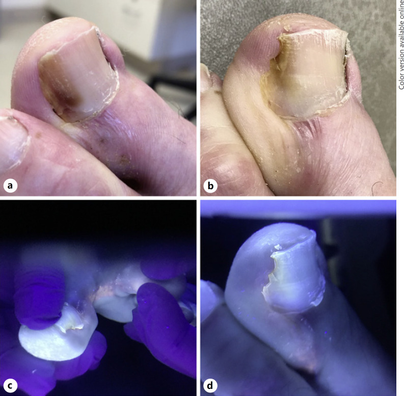
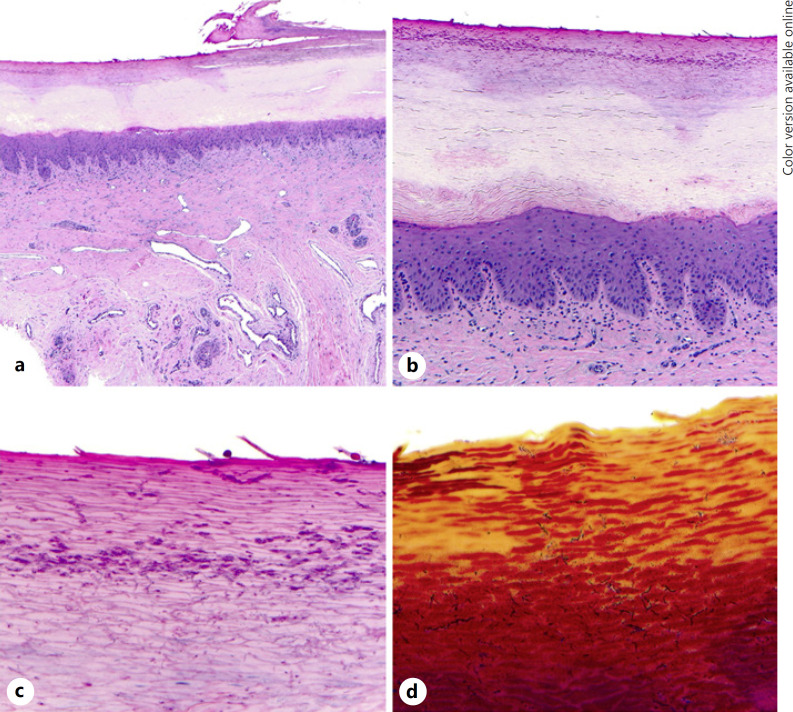
Nail pathology may reflect a wide array of localized and systemic dermatological conditions. Certain nail findings such as melanonychia can create diagnostic challenges even to nail experts. We report a case of a 78-year-old man who presented with melanonychia of the great toe. Nail clipping showed focal melanin deposition, and dermoscopy demonstrated a region of localized erythema in the lunula concerning for possible melanocytic neoplasm. Subsequent nail biopsy showed numerous vertically oriented filamentous bacteria and coccobacilli within the nail plate consistent with a diagnosis of subungual nail erythrasma. Nail erythrasma is a rare entity. Additionally, this case highlights a new clinical presentation of nail erythrasma as melanonychia.
Keywords: Cornyebacteria; Erythrasma; Nail matrix; Nail plate.
Copyright © 2020 by S. Karger AG, Basel.
Conflict of interest statement
The authors declare no conflict of interest.
Figures
References
-
- Negroni P. [Erythrasma of the nails] Med Cutan Ibero Lat Am. 1976;4((5)):349–57. - PubMed
-
- Duhard E, Calvet C, Mariotte N, Tichet J, Vaillant L. [Prevalence of longitudinal melanonychia in the white population] Ann Dermatol Venereol. 1995;122((9)):586–90. - PubMed
-
- Jellinek N. Nail matrix biopsy of longitudinal melanonychia: diagnostic algorithm including the matrix shave biopsy. J Am Acad Dermatol. 2007;56((5)):803–10. - PubMed
-
- Haneke E, Baran R. Longitudinal melanonychia. Dermatol Surg. 2001;27((6)):580–4. - PubMed
-
- Sommer LL, Reboli AC, Heymann WR. Bacterial diseases. In: Bolognia JL, Schaffer JV, Cerroni L, editors. Dermatology. 4th ed. Vol. 1. Philadelphia: Elsevier; 2018. pp. p.1274–7.
Publication types
LinkOut - more resources
Full Text Sources
