

Subungual Elastofibroma
- PMID: 33614720
- PMCID: PMC7879278
- DOI: 10.1159/000510857
Subungual Elastofibroma
Abstract
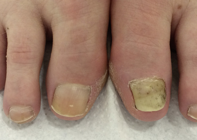
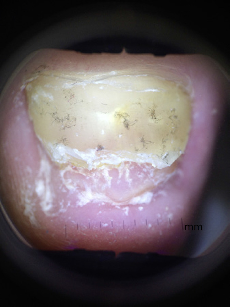
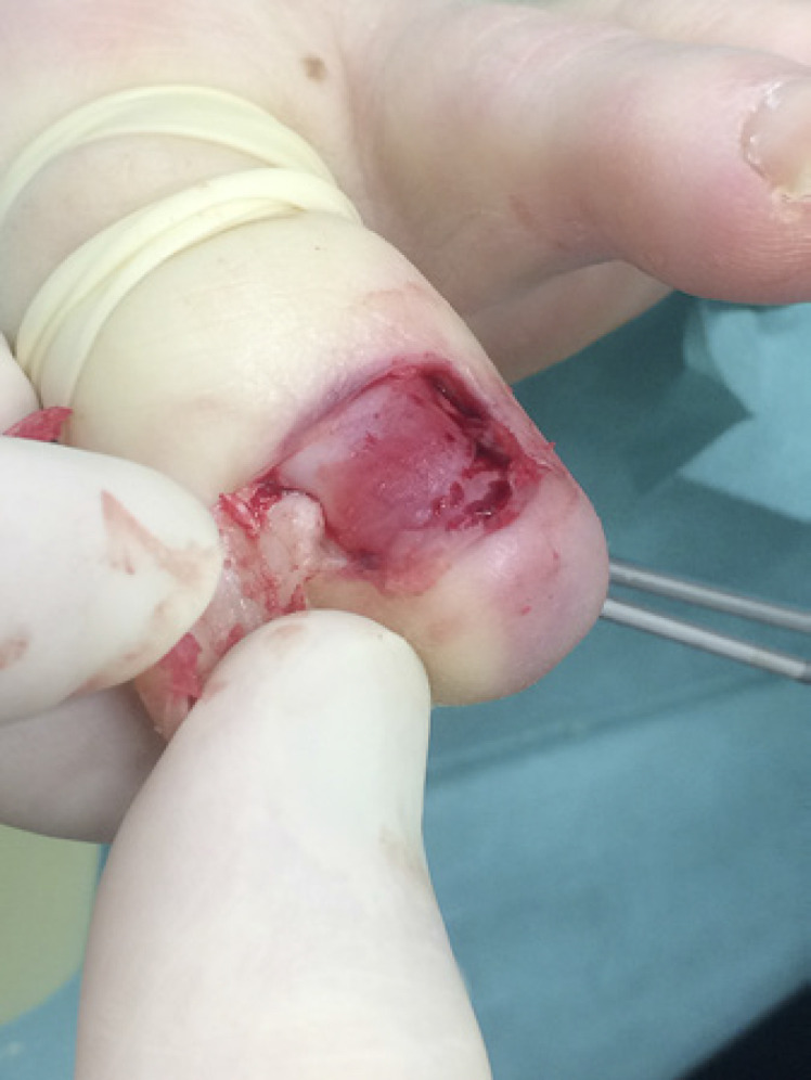
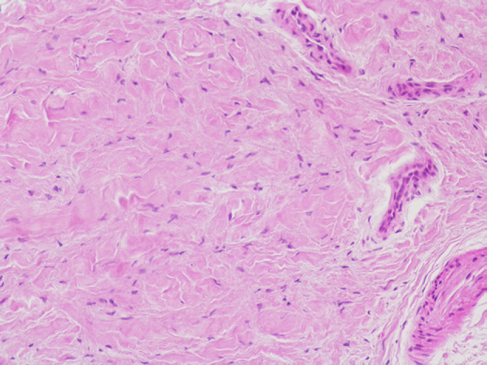
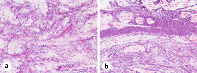
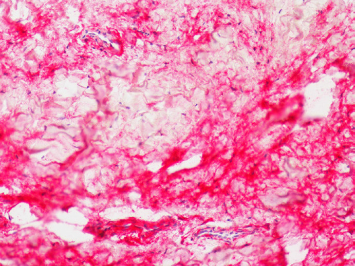
Cutaneous elastofibroma is part of the connective tissue nevus complex. Two subungual cases remotely similar to the one presented here have been described before. This patient presented with an unusual form of subungual elastofibroma of the big toe, which was surgically removed. Histopathology revealed a connective tissue tumor extremely rich in very fine elastic fibers. Their relationship to oxytalan and elaunin fibers is discussed as is the potential association of this nail bed lesion with the onychodermis.
Keywords: Connective tissue nevi; Nail tumors; Onychodermis; Onychopathology; Subungual elastofibroma.
Copyright © 2020 by S. Karger AG, Basel.
Conflict of interest statement
The authors have no conflicts of interest to disclose.
Figures
Similar articles
-
Light and electron microscopic study on the oxytalan elaunin and elastic fibers in the inferior segment of the human esophagus.Anat Anz. 1982;152(2):141-57. Anat Anz. 1982. PMID: 7158796
-
Elastic system fibers (oxytalan, elaunin and elastic fibers) in the skin of a freshwater teleost : optical and electron microscopy study.Arch Anat Microsc Morphol Exp. 1980;69(4):259-66. Arch Anat Microsc Morphol Exp. 1980. PMID: 7212696
-
Tumors of the nail unit. A review. Part II: acquired localized longitudinal pachyonychia and masked nail tumors.Am J Dermatopathol. 2013 Oct;35(7):693-709; quiz 710-2. doi: 10.1097/DAD.0b013e318293f387. Am J Dermatopathol. 2013. PMID: 24056180 Review.
-
Onycholemmal Horn: An Exceedingly Rare Subungual Tumor.Skin Appendage Disord. 2021 Aug;7(5):413-417. doi: 10.1159/000516303. Epub 2021 Jun 1. Skin Appendage Disord. 2021. PMID: 34604335 Free PMC article.
-
Metastatic tumors to the nail unit: subungual metastases.Dermatol Surg. 2001 Mar;27(3):280-93. Dermatol Surg. 2001. PMID: 11277898 Review.
Cited by
-
Rare rectal elastofibroma: diagnostic challenges and case report.Discov Oncol. 2025 May 19;16(1):815. doi: 10.1007/s12672-025-02556-6. Discov Oncol. 2025. PMID: 40388002 Free PMC article.
References
-
- Aroni K, Aivaliotis M, Davaris P. Isolated connective tissue nevus originating subungually: report of a unique case. J Dermatol. 2001;28((12)):765–6. - PubMed
-
- Wolner ZJ, Liebman TN, Lowenstein EJ. Acquired elastoma in subungual location. Dermatol Online J. 2017;23((9)):11. - PubMed
-
- Buschke A, Ollendorff H. Ein Fall von Dermatofibrosis lenticularis disseminata und Osteopathia condensans disseminata. Dermatol Wschr. 1928;86:257–62.
-
- Weidman FD, Anderson NP, Ayres S. Juvenile elastoma. Arch Dermatol. 1933;28((2)):182–9.
-
- Saussine A, Marrou K, Delanoé P, Bodak N, Hamel D, Picard A, et al. Connective tissue nevi: an entity revisited. J Am Acad Dermatol. 2012;67((2)):233–9. - PubMed
Publication types
LinkOut - more resources
Full Text Sources
