. 2021 Feb 4:8:622189.
doi: 10.3389/fvets.2021.622189. eCollection 2021.

Detection of Epithelial Cell Adhesion Molecule in Feline Normal and Tumor Cell Lines and Tissues With Selected Commercial Anti-human EpCAM Antibodies

Affiliations

Detection of Epithelial Cell Adhesion Molecule in Feline Normal and Tumor Cell Lines and Tissues With Selected Commercial Anti-human EpCAM Antibodies

Christa Y Heyward et al. Front Vet Sci. .

Abstract

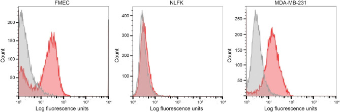
Epithelial cell adhesion molecule (EpCAM) is a transmembrane protein expressed at intercellular junctions in epithelial cells. As an epithelial biomarker, it used for immunologic-based capture of epithelial-derived circulating tumor cells (CTCs) in human patients with different carcinomas. EpCAM expression has not been described in normal or neoplastic epithelial tissues in cats. Our goal was to find a commercial antibody that recognizes surface EpCAM expression for CTC detection. We tested two anti-human EpCAM antibodies, designated for use with flow cytometry, for detection of surface EpCAM expression on feline cell lines derived from normal mammary and renal epithelia and mammary and oropharyngeal squamous cell carcinomas in cats. Only one of the antibodies, a goat polyclonal antibody, labeled normal and neoplastic feline mammary epithelial cells and oropharyngeal squamous cell carcinoma cells; no labeling was observed for normal feline kidney epithelial cells. At low dilution, this antibody immunohistochemically stained the intercellular junctions of normal pancreatic, intestinal and mammary epithelium, as well as neoplastic mammary epithelium in feline tissues; however, oral mucosa, skin, and an oropharyngeal squamous cell carcinoma showed no positive immunostaining. The antibody only weakly bound feline squamous cell carcinoma cell lines under static adhesion. Our results indicate that EpCAM is expressed in specific epithelia in cats but is variably expressed in feline mammary tumors and oropharyngeal squamous cell carcinoma. A higher avidity cross-reactive or feline-specific antibody will be required to further investigate EpCAM expression in normal and neoplastic feline tissue or for detecting CTCs in the blood of tumor-bearing cats.

Keywords: TROP-1/Ep-CAM; cancer; cat; circulating tumor cells; flow cytometry; immunohistochemistry; mammary carcinoma; squamous cell carcinoma.


Conflict of interest statement

The authors declare that the research was conducted in the absence of any commercial or financial relationships that could be construed as a potential conflict of interest.

Figures

Figure 1
Figure 1
Alignment of human and feline EpCAM amino acid sequences using UniProt sequences for human (P16422) and feline (M3WIV4) EpCAM sequences (Jalview, version 2.11.1.0). Similar colored boxes show sequence alignment.
Figure 2
Figure 2
Binding of the two selected anti-human EpCAM antibodies (SB EpCAM, R&D EpCAM) to human breast carcinoma cell lines, MCF-7 and MDA-MB-231, by flow cytometry. Representative frequency distribution curves of logarithmic fluorescent intensity for each cell line are shown (n = 3 independent experiments) (EpCAM antibodies: pink solid curves; rabbit serum or goat gamma globulin controls: gray solid curves with dotted line).
Figure 3
Figure 3
Labeling of feline normal mammary (FMEC) and renal (NLFK) epithelial cell lines with a goat polyclonal anti-human EpCAM antibody (R&D EpCAM) with flow cytometry, with a human breast cancer cell line (MDA-MB-231) as a positive control. Representative frequency distribution curves of logarithmic fluorescent intensity for each cell line are shown (EpCAM antibodies: pink solid curves; rabbit serum or goat gamma globulin controls: gray solid curves) (n ≥ 3 independent experiments).
Figure 4
Figure 4
Screening of feline mammary carcinoma (CAT-MT, K12-72.1), oral squamous cell carcinoma (SCCF-2, SCCF-3), and an injection site sarcoma-derived (C10) cell lines with the rabbit monoclonal (SB EpCAM) and goat polyclonal (R&D EpCAM) anti-human EpCAM antibodies with flow cytometry. The MCF-7 human breast carcinoma cell line was used as a positive control. Representative frequency distribution curves of logarithmic fluorescent intensity for each cell line are shown (n ≥ 3 independent experiments). Only the R&D EpCAM antibody showed consistent binding to the feline epithelial cell lines but not the sarcoma cell line (EpCAM antibodies: pink solid curves; rabbit serum or goat gamma globulin controls: gray solid curves with dotted line).
Figure 5
Figure 5
Recombinant human EpCAM competes with tumor cells for binding to the R&D goat polyclonal anti-EpCAM antibody on flow cytometric analysis. Addition of 0.5 or 1.5 μg recombinant EpCAM caused a dose-dependent decrease in binding of the R&D EpCAM antibody to the MCF-7 human breast carcinoma cell line (A). Exposure to 0.5 μg of recombinant protein decreased binding, whereas the higher concentration of 1.5 μg abolished binding, of the R&D EpCAM antibody to the feline oral squamous cell carcinoma cell line, SCCF-3 (B). The recombinant protein did not influence background fluorescence of the goat gamma globulin control for either cell line.
Figure 6
Figure 6
Immunohistochemical staining of duodenum (A,B) and pancreatic epithelium (C,D) with the R&D goat polyclonal EpCAM antibody (A,C) and a goat IgG control (B,D) using an initial non-optimized staining protocol on a feline tissue array (DAB chromogen). Positive membranous staining, outlining the intercellular junctions of epithelial cells, is evident with the R&D EpCAM antibody in the duodenum (A) and pancreas (C). In the pancreas, both acinar (C) and ductular epithelium (not shown) showed similar membrane staining. Substantial background cytoplasmic staining was evident with the negative goat IgG control, but staining of the cell membranes was lacking in either the duodenum (B) or pancreas (D). Scale bar = 20 μm.
Figure 7
Figure 7
Immunohistochemical staining of a grade III mammary tubular carcinoma in a cat with the R&D goat polyclonal anti-EpCAM antibody using the optimized protocol (DAB chromogen). Several areas of the section contained unaffected normal mammary epithelium, which showed positive cell membrane staining (A). Only small sections of the tumor showed positive membrane staining (B), whereas much of the tumor did not stain with the antibody (C). Minimal staining was seen with the goat IgG control in the normal mammary epithelium (not shown) or throughout the mammary tumor (D, scale bar = 20 μm).
Figure 8
Figure 8
Binding of feline oral squamous cell carcinoma cell lines to the R&D goat polyclonal anti-EpCAM antibody-coated coverslips using a static adhesion assay. Binding was determined by a blinded observer, who counted fluorescent nuclei in 10 randomly photographed fields per coverslip, after cells were allowed to bind for 1 h, followed by washing and fluorescent labeling of nuclei. Individual data points are shown with medians (red line). We tested the feline oral squamous cell carcinoma cell lines (SCCF-2, A, and SCCF-3, B) with the MCF-7 human mammary carcinoma (C) and feline C10 injection site sarcoma (D) cell lines as positive and negative cell controls, respectively, on R&D EpCAM-coated coverslips (triangles), with goat gamma globulin (γ-globulin; squares) and the rabbit monoclonal SB EpCAM antibody (inverted triangle) as negative coating controls for the feline cell lines. The SB EpCAM antibody was used as a positive control for the MCF-7 cells. Fibronectin was used as an integrin-mediated binding control (circles). Note that there is a different scale on the Y-axis for each cell line. *p < 0.05 comparing R&D EpCAM- to goat γ-globulin-coated coverslips. **p < 0.05 comparing R&D EpCAM- to SB EpCAM-coated coverslips.

Similar articles

Cited by

References

    1. Mader S, Pantel K. Liquid biopsy: current status and future perspectives. Oncol Res Treat. (2017) 40:404–8. 10.1159/000478018 - DOI - PubMed
    1. Wang Y, Yu X, Hartmann D, Zhou J. Circulating tumor cells in peripheral blood of pancreatic cancer patients and their prognostic role: a systematic review and meta-analysis. HPB (Oxford). (2020) 22:660–9. 10.1016/j.hpb.2019.11.003 - DOI - PubMed
    1. Jia M, Mao Y, Wu C, Wang S, Zhang H. A platform for primary tumor origin identification of circulating tumor cells via antibody cocktail-based in vivo capture and specific aptamer-based multicolor fluorescence imaging strategy. Anal Chim Acta. (2019) 1082:136–45. 10.1016/j.aca.2019.07.051 - DOI - PubMed
    1. Han L, Li J, Zhang W, Song P, Li H, Li S. Clinical significance of tumor cells in the peripheral blood of patients with esophageal squamous cell carcinoma. Medicine. (2019) 98:e13921. 10.1097/MD.0000000000013921 - DOI - PMC - PubMed
    1. Huebner H, Fasching PA, Gumbrecht W, Jud S, Rauh C, Matzas M, et al. . Filtration based assessment of CTCs and CellSearch® based assessment are both powerful predictors of prognosis for metastatic breast cancer patients. BMC Cancer. (2018) 18:204. 10.1186/s12885-018-4115-1 - DOI - PMC - PubMed