

Vinblastine treatment decreases the undifferentiated cell contamination of human iPSC-derived intestinal epithelial-like cells
- PMID: 33614822
- PMCID: PMC7868938
- DOI: 10.1016/j.omtm.2021.01.005
Vinblastine treatment decreases the undifferentiated cell contamination of human iPSC-derived intestinal epithelial-like cells
Abstract
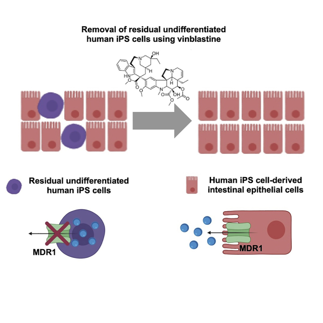
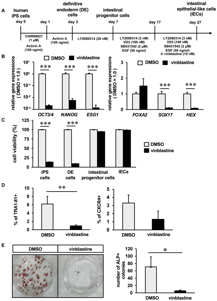
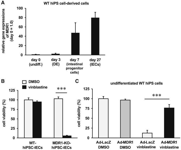
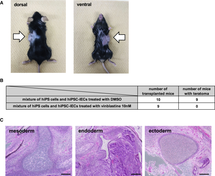
Human induced pluripotent stem cell-derived intestinal epithelial cells (hiPSC-IECs) are expected to be utilized in regenerative medicine. To perform a safe transplantation without the risk of tumor formation, residual undifferentiated hiPSCs must be removed from hiPSC-IECs. In this study, we examined whether vinblastine (a multiple drug resistance 1 [MDR1] substrate) could remove residual undifferentiated hiPSCs in hiPSC-IECs and attempted to generate hiPSC-IECs applicable to transplantation medicine. We found that the expression levels of pluripotent markers were largely decreased and those of intestinal markers were increased by vinblastine treatment. The treatment of undifferentiated hiPSCs with vinblastine significantly decreased their viability. These results suggested that undifferentiated hiPSCs can be eliminated from hiPSC-IECs by vinblastine treatment. We hypothesized that MDR1-negative cells (such as undifferentiated hiPSCs) die upon vinblastine treatment because they are unable to excrete vinblastine. As expected, the cell viability of MDR1-knockout hiPSC-IECs was significantly decreased by vinblastine treatment. Furthermore, teratomas were formed by subcutaneous transplantation of hiPSC-IECs mixed with undifferentiated hiPSCs into mice, but they were not observed when the transplanted cells were pre-treated with vinblastine. Vinblastine-treated hiPSC-IECs would be an effective cell source for safe regenerative medicine.
Keywords: differentiation; iPS cell; intestinal epithelial cells; regenerative medicine; vinblastine.
© 2021 The Author(s).
Conflict of interest statement
We receive research grants from Takara Bio.
Figures

Similar articles
-
Generation of intestinal organoids derived from human pluripotent stem cells for drug testing.Sci Rep. 2020 Apr 6;10(1):5989. doi: 10.1038/s41598-020-63151-z. Sci Rep. 2020. PMID: 32249832 Free PMC article.
-
Plasma-activated medium selectively eliminates undifferentiated human induced pluripotent stem cells.Regen Ther. 2016 Sep 10;5:55-63. doi: 10.1016/j.reth.2016.07.001. eCollection 2016 Dec. Regen Ther. 2016. PMID: 31245502 Free PMC article.
-
Tumorigenicity-associated characteristics of human iPS cell lines.PLoS One. 2018 Oct 4;13(10):e0205022. doi: 10.1371/journal.pone.0205022. eCollection 2018. PLoS One. 2018. PMID: 30286143 Free PMC article.
-
[In vitro tumorigenicity tests for process control of health care products derived from human induced pluripotent stem cells].Yakugaku Zasshi. 2013;133(12):1381-8. doi: 10.1248/yakushi.13-00232-3. Yakugaku Zasshi. 2013. PMID: 24292187 Review. Japanese.
-
Maintenance of an undifferentiated state of human induced pluripotent stem cells through migration-dependent regulation of the balance between cell-cell and cell-substrate interactions.J Biosci Bioeng. 2015 Jun;119(6):617-22. doi: 10.1016/j.jbiosc.2014.10.024. Epub 2014 Nov 22. J Biosci Bioeng. 2015. PMID: 25468424 Review.
Cited by
-
Pharmacological potential of bioactive compounds in Catharanthus roseus extract: A comprehensive review.Toxicol Rep. 2025 Mar 17;14:101998. doi: 10.1016/j.toxrep.2025.101998. eCollection 2025 Jun. Toxicol Rep. 2025. PMID: 40213418 Free PMC article. Review.
-
Generation of Caco-2 cells stably expressing CYP3A4·POR·UGT1A1 and CYP3A4·POR·UGT1A1*6 using a PITCh system.Arch Toxicol. 2022 Feb;96(2):499-510. doi: 10.1007/s00204-021-03175-0. Epub 2021 Oct 16. Arch Toxicol. 2022. PMID: 34654938
-
Functional intestinal monolayers from organoids derived from human iPS cells for drug discovery research.Stem Cell Res Ther. 2024 Feb 29;15(1):57. doi: 10.1186/s13287-024-03685-5. Stem Cell Res Ther. 2024. PMID: 38424603 Free PMC article.
References
-
- Iwao T., Kodama N., Kondo Y., Kabeya T., Nakamura K., Horikawa T., Niwa T., Kurose K., Matsunaga T. Generation of enterocyte-like cells with pharmacokinetic functions from human induced pluripotent stem cells using small-molecule compounds. Drug Metab. Dispos. 2015;43:603–610. - PubMed
LinkOut - more resources
Full Text Sources
Other Literature Sources
Research Materials
