. 2020 Nov 10;6(2):472-483.
doi: 10.1016/j.ekir.2020.10.040. eCollection 2021 Feb.

Recessive Mutations in SYNPO2 as a Candidate of Monogenic Nephrotic Syndrome

Affiliations

Recessive Mutations in SYNPO2 as a Candidate of Monogenic Nephrotic Syndrome

Youying Mao et al. Kidney Int Rep. .

Abstract

Introduction: Most of the approximately 60 genes that if mutated cause steroid-resistant nephrotic syndrome (SRNS) are highly expressed in the glomerular podocyte, rendering SRNS a "podocytopathy."

Methods: We performed whole-exome sequencing (WES) in 1200 nephrotic syndrome (NS) patients.

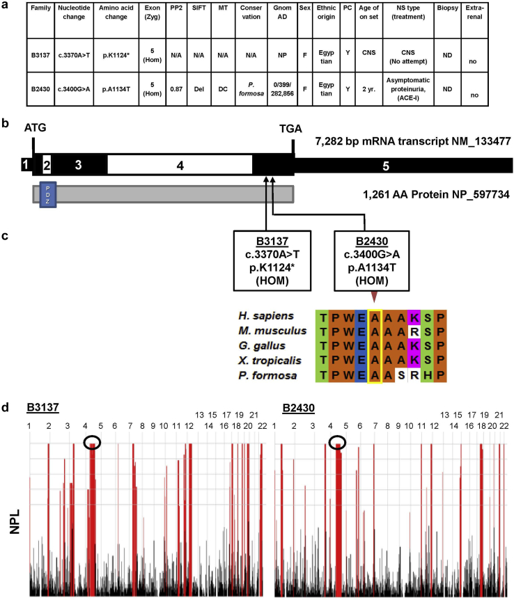
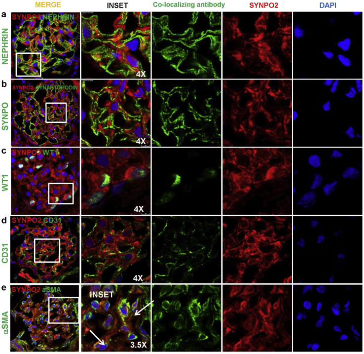
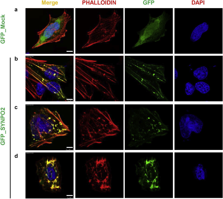
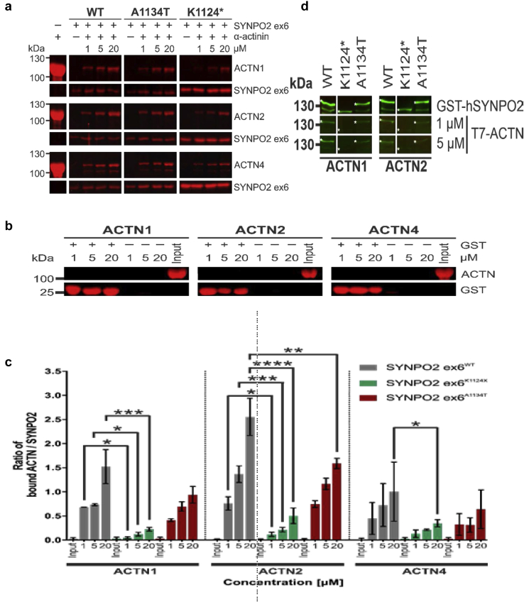
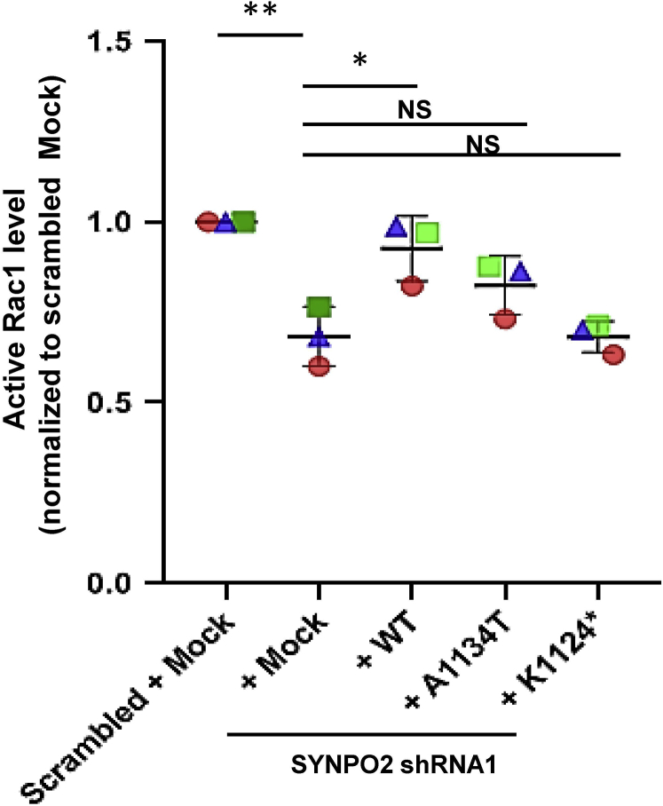
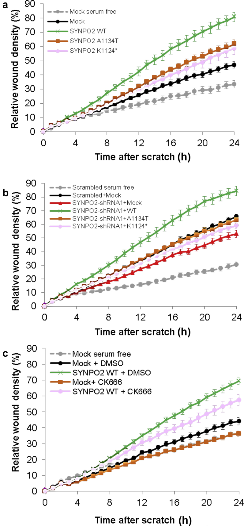
Results: We discovered homozygous truncating and homozygous missense mutation in SYNPO2 (synaptopodin-2) (p.Lys1124∗ and p.Ala1134Thr) in 2 patients with childhood-onset NS. We found SYNPO2 expression in both podocytes and mesangial cells; however, notably, immunofluorescence staining of adult human and rat kidney cryosections indicated that SYNPO2 is localized mainly in mesangial cells. Subcellular localization studies reveal that in these cells SYNPO2 partially co-localizes with α-actinin and filamin A-containing F-actin filaments. Upon transfection in mesangial cells or podocytes, EGFP-SYNPO2 co-localized with α-actinin-4, which gene is mutated in autosomal dominant SRNS in humans. SYNPO2 overexpression increases mesangial cell migration rate (MMR), whereas shRNA knockdown reduces MMR. Decreased MMR was rescued by transfection of wild-type mouse Synpo2 cDNA but only partially by cDNA representing mutations from the NS patients. The increased mesangial cell migration rate (MMR) by SYNPO2 overexpression was inhibited by ARP complex inhibitor CK666. SYNPO2 shRNA knockdown in podocytes decreased active Rac1, which was rescued by transfection of wild-type SYNPO2 cDNA but not by cDNA representing any of the 2 mutant variants.

Conclusion: We show that SYNPO2 variants may lead to Rac1-ARP3 dysregulation, and may play a role in the pathogenesis of nephrotic syndrome.

Keywords: SYNPO2; monogenic kidney disease; nephrotic syndrome.


Figures

Figure 1
Figure 1
SYNPO2 mutations identified in 2 families in nephrotic syndrome (NS). (a) Summary of genetic and phenotypic data for 2 patients with SYNPO2 mutations. ACE-I, angiotensin-converting enzyme inhibitor; CNS, congenital nephrotic syndrome; DC, disease causing; Del, deleterious; F, female; gnomAD, Genome Aggregation database; Hom, homozygous; M, male; MT, mutation taster; N/A, not applicable; ND, not done; NP, not present in control variant database; PC, parental consanguinity; PP2, PolyPhen-2 prediction score; SIFT, “Sorting Tolerant from Intolerant” prediction score; Zyg, Zygosity. (b) Exon structure (upper bar) and protein domain content (lower bar) structures of SYNPO2 are shown with arrows indicating positions of mutations in patients (B3137 and B2430) with NS or persistent proteinuria. (c) Evolutionary conservation of amino acid position A1134 in SYNPO2 protein across evolution. (d) Homozygosity mapping data across the genome was generated using nonparametric LOD scores (NPL scores) based on WES variant data using Homozygosity Mapper for individuals B3137 and B2430. Black circles demonstrate the NPL peak regions, in which SYNPO2 mutations were positioned.
Figure 2
Figure 2
SYNPO2 immunofluorescence stain with antibody (Ab#1 Abcam, ab50192) in rat glomeruli. Adult rat kidney sections were stained with SYNPO2, and co-stained with cell type marker antibodies against nephrin (podocyte slit membrane), SYNPO (podocyte cytoplasm), WT1 (podocyte nucleus), CD31 (endothelial cell), and aSMA (mesangial cells). SYNPO2 staining is detected in rat glomeruli by immunofluorescence on kidney frozen sections but is not co-localizing with nephrin, SYNAPTOPODIN, WT1, or CD31. However, SYNPO2 partially co-localizes with aSMA, a mesangial cell marker (white arrows). DAPI stains nuclear (blue). Bar = 5 μm.
Figure 3
Figure 3
SYNPO2 co-localizes with F-actin networks in 2 distinct patterns in mesangial cells. (a) Rat mesangial cells (RMCs) transfected with green fluorescent protein (GFP) mock negative control were almost devoid of large actin fibers in the cell body but displayed strong F-actin staining around the cell periphery. (b, c) Transfection of RMCs with GFP_SYNPO2. SYNPO2 induces 2 distinct F-actin patterns, co-localizing with these networks. F-actin staining shows long and well-organized actin bundles, frequently oriented parallel along the axis of the cell. (d) The alternative pattern shows perinuclear and bipolar fusiform morphology with thick actin irregular bundles. Bar = 5 μm.
Figure 4
Figure 4
Mutations in SYNPO2 reduce binding to α-actinin. (a) Pulldown experiments. Different quantities of α-actinin (ACTN) were added to GST-SYNPO2 ex6 (aminoacids 1085−1261) variants bound to GSH-beads. Gel fractions show bound ACTN1, 2, and 4 (upper panels), as well as GSH-bound SYNPO2 (lower panels). Left panels show α-actinin for quality control. Concentrations (μM) correspond to amount of actinin input. (b) Control pulldown experiments. No nonspecific binding of α-actinin to GSH-beads (−) or GST-coupled beads (+) could be observed. (c) Quantification of the experiment shown in (a). Ratios of α-actinin (ACTN) to SYNPO2 bands were calculated to quantify bound α-actinin-1, α-actinin-2, and α-actinin-4. ACTN1, ACTN2, and ACTN4 show significantly impaired binding to SYNPO2K1124∗ mutants compared to wild-type SYNPO2. The A1134T-mutation significantly impairs binding of α-actinin2 at higher concentrations. Bars represent standard error of n = 3. ∗P < 0.05; ∗∗P < 0.01; ∗∗∗P < 0.001; ∗∗∗∗P < 0.0001. (d) Western blot overlay of α-actinin1, 2 to immobilized SYNPO2. Purified WT or mutant SYNPO2 (amino acids 396−1261) are indicated with asterisks. In comparison to wild type, binding of α-actinin1, and 2 is impaired for both SYNPO2 mutants.
Figure 5
Figure 5
Synpo2 rescued active Rac1 in SYNPO2 knockdown cell line, but not by mutants from nephrotic syndrome (NS) patients. Active levels of Rac1 were measured by Rac1 G-LISA assay. shRNA-mediated knockdown of SYNPO2 in human podocytes (+ Mock) reduced active Rac1. Overexpression of wild-type Synpo2 cDNA (+ WT) rescued this effect, but the Synpo2 cDNA constructs reflecting mutations in NS patients, failed to rescue reduction of active Rac1 (+A1134T, +K1124∗). P values calculated by one-way analysis of variance. ∗P < 0.05, ∗∗P < 0.01. NS, nonsignificant.
Figure 6
Figure 6
Wild-type but not mutant SYNPO2 increases migration rate and acts upstream of ARP2/3. (a) In a human mesangial cell line expressing Mock negative control, MMR is increased following serum addition (black) relative to serum-free conditions (gray). All subsequent experiments were performed in serum replete conditions. Overexpression of SYNPO2 WT (green) strongly increased MMR compared to Mock (black). Human SYNPO2 cDNA constructs representing mutants from NS patients (p.A1134T and p. K1124∗) partially rescued MMR compared to Mock (orange and pink). (B) In a human mesangial cell line expressing scrambled shRNA, MMR is increased following serum addition (black) relative to serum-free conditions (gray). All subsequent experiments were performed in serum-replete conditions. Knockdown of SYNPO2 (red) showed reduced MMR compared with scrambled shRNA with Mock overexpression (black). Migration was rescued by overexpression of WT Synpo2 construct (green). Mouse Synpo2 cDNA constructs reflecting mutations in NS patients only partially rescued the MMR (orange and pink). (c) ARP3 complex is an effector for mesangial cell migration downstream of SYNPO2. In a human mesangial cell line expressing Mock negative control, MMR is increased following serum addition (black) with dimethylsulfoxide (DMSO) control relative to serum-free conditions (gray). All subsequent experiments were performed in serum-replete conditions. Overexpression of SYNPO2 WT (green) with DMSO control strongly increased MMR compared to Mock (black) with DMSO control. However, the increased migration (green) was reduced with ARP3 inhibitor CK666 (pink). In Mock transfected cells, migration was also reduced with CK666 (orange) compared to DMSO (black). CK666 = 50 μM.

References

    1. Wiggins R.C. The spectrum of podocytopathies: a unifying view of glomerular diseases. Kidney Int. 2007;71:1205–1214. - PubMed
    1. Trautmann A., Schnaidt S., Lipska-Zietkiewicz B.S. Long-term outcome of steroid-resistant nephrotic syndrome in children. J Am Soc Nephrol. 2017;28:3055–3065. - PMC - PubMed
    1. Sadowski C.E., Lovric S., Ashraf S. A single-gene cause in 29.5% of cases of steroid-resistant nephrotic syndrome. J Am Soc Nephrol. 2015;26:1279–1289. - PMC - PubMed
    1. Warejko J.K., Tan W., Daga A. Whole exome sequencing of patients with steroid-resistant nephrotic syndrome. Clin J Am Soc Nephrol. 2018;13:53–62. - PMC - PubMed
    1. Park J., Shrestha R., Qiu C. Single-cell transcriptomics of the mouse kidney reveals potential cellular targets of kidney disease. Science. 2018;360:758–763. - PMC - PubMed