. 2021 Feb 9;2(1):100334.
doi: 10.1016/j.xpro.2021.100334. eCollection 2021 Mar 19.

Massive expansion and cryopreservation of functional human induced pluripotent stem cell-derived cardiomyocytes

Affiliations

Massive expansion and cryopreservation of functional human induced pluripotent stem cell-derived cardiomyocytes

Renee G C Maas et al. STAR Protoc. .

Erratum in

Abstract

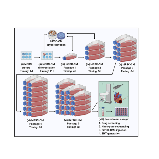
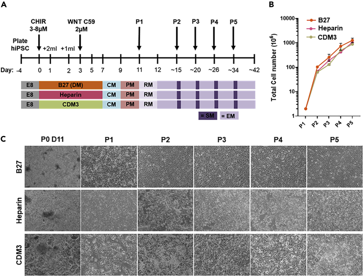
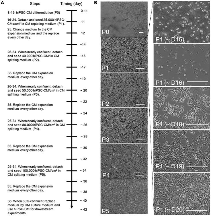
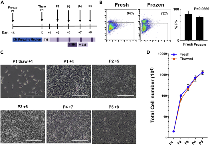
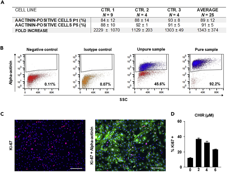
Since the discovery of human induced pluripotent stem cells (hiPSCs), numerous strategies have been established to efficiently derive cardiomyocytes from hiPSCs (hiPSC-CMs). Here, we describe a cost-effective strategy for the subsequent massive expansion (>250-fold) of high-purity hiPSC-CMs relying on two aspects: removal of cell-cell contacts and small-molecule inhibition with CHIR99021. The protocol maintains CM functionality, allows cryopreservation, and the cells can be used in downstream assays such as disease modeling, drug and toxicity screening, and cell therapy. For complete details on the use and execution of this protocol, please refer to Buikema (2020).

Keywords: Cell culture; Cell differentiation; Stem cells.


Conflict of interest statement

J.W.B. and S.M.W. have filed for a patent with the US Patent and Trademark Office regarding the effect of bioactive lipids plus Wnt signaling activation on hiPSC-CM proliferation/expansion.

Figures

None
Graphical abstract
Figure 1
Figure 1
Massive expansion of hiPSC-CMs from various differentiation methods (A) Schematic representation of Wnt-based directed cardiac differentiation and subsequent expansion of hiPSC-CMs generated by three different medium compositions ((B27; Lian, 2012; Heparin; Lin, 2017, and CDM3; Burridge, 2014). Timeline indicating B27-medium with insulin and additional CHIR99021 (CHIR) usage required for expansion. CM, CM culture medium; PM, CM purification medium; RM, CM replating medium; EM, CM expansion medium; SM, CM splitting medium; P, passage. (B) Graph displaying the hiPSC-CM expansion capacity from hiPSC-CMs generated by the indicated differentiation method. One hiPSC line was used for these experiments. n = 7, n = 2, n = 2 respectively from P0 day 11 to P5. Data are represented as mean ± SD. (C) Bright-field images at different time points of hiPSC-CM expansion with the input of the indicated differentiation methods. Scale bar, 200 μm.
Figure 2
Figure 2
Visualized timeline of hiPSC-CM expansion (A) Timeline diagram displaying the steps required for expansion and passaging of hiPSC-CMs. (B) Time-lapse images of cell morphology at the indicated passage numbers and/or culture days. Left: Images of cell confluency on day of passaging for P0-5. Right: Daily consecutive images of P1 hiPSC-CMs demonstrating cell seeding density and growth speed between P1 and P2. Replating at day 15, start of CHIR9021 (CHIR) treatment at day 16, cell cluster formation at day 14–19 is observed the days after and a nearly confluent monolayer before passaging at D18–20. Scale bar, 200 μm. D, day; P, passage.
Figure 3
Figure 3
Cryopreservation and subsequent expansion of beating hiPSC-CMs (A) Schematic representation of small-molecule-based Wnt-modulated directed cardiac differentiation and subsequent cryopreservation and expansion. hiPSC-CMs can be stored in liquid nitrogen after passage 1–2 and after thawing expanded for 5 passages (P) before downstream assays. TM, CM thawing medium; EM, CM expansion medium; SM, CM splitting medium; P, passage. (B) Left panels are flow cytometer detection of dead cells with propidium iodide (PI) after detaching or after thawing of cardiomyocytes, cryopreserved on day 15. Right is the quantification of fresh versus thawed PI % (n = 5 per condition, p < 0.05, paired t test.). Data are represented as mean ± SD. (C) Representative images of the thawed hiPSC-CMs before starting the expansion protocol and during passaging (scale bar, 100 μm). (D) Quantification of cardiomyocyte expansion curve of fresh versus thawed hiPSC-CMs, n = 7, n = 5 respectively from thawed P1 to P5. Data are represented as mean ± SD.
Figure 4
Figure 4
Differentiation efficiency and expansion capacity in different control hiPSC lines (A) Quantitative analysis of small-molecule-based Wnt-modulated directed cardiac differentiation at the passage (P) number P1 and P5. Replicate numbers refer to the number of performed expansion experiments from individual differentiation batches. The percentages of increase in hiPSC-CMs are relative to the input of day 11 hiPSC-CMs. Mean values ± SD are given. (B) Representative gating strategy for α-actinin positive hiPSC-CMs in a pure population versus negative control, isotype control, and an impure or un-purified hiPSC-CM culture. The number of α-actinin positive analyzed cells is 25 × 105. SSC, side scatter. (C) Representative immunofluorescence for proliferation assessed by Ki67 expression after 48 h of CHIR treatment. Immunofluorescence: Hoechst (blue), Ki67 (red), and α-actinin (green). Scale bar, 200 μm. (D) Quantitative graph indicates high proliferation (37%) of cardiomyocytes stimulated with the optimal dose of 2 μM CHIR.

References

    1. Buikema J.W. Wnt activation and reduced cell-cell contact synergistically induce massive expansion of functional human iPSC-derived cardiomyocytes. Cell Stem Cell. 2020;1:50–63. - PMC - PubMed
    1. Burridge P. Chemically defined generation of human cardiomyocytes. Nat. Methods. 2014;8:855–860. - PMC - PubMed
    1. Lian X. Robust cardiomyocyte differentiation from human pluripotent stem cells via temporal modulation of canonical wnt signaling. Proc. Natl. Acad. Sci. U S A. 2012;109:1848–1857. - PMC - PubMed
    1. Lin Y. Heparin promotes cardiac differentiation of human pluripotent stem cells in chemically defined albumin-free medium, enabling consistent manufacture of cardiomyocytes. Stem Cells Transl. Med. 2017;6:527–538. - PMC - PubMed

Publication types

MeSH terms