

Increased PD-L1 Expression in Human Skin Acutely and Chronically Exposed to UV Irradiation
- PMID: 33615483
- PMCID: PMC8277687
- DOI: 10.1111/php.13406
Increased PD-L1 Expression in Human Skin Acutely and Chronically Exposed to UV Irradiation
Abstract
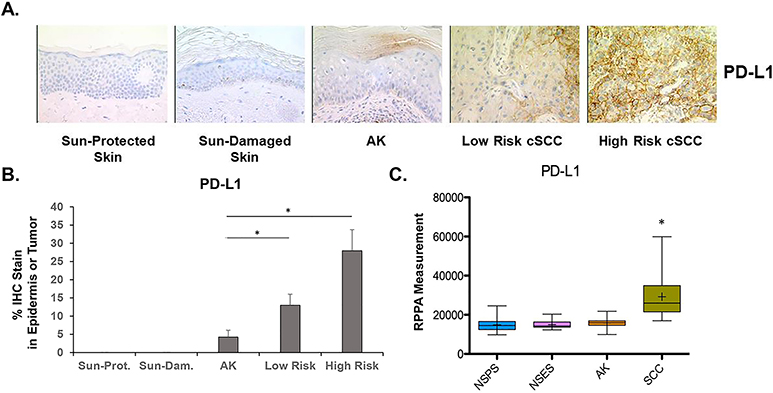
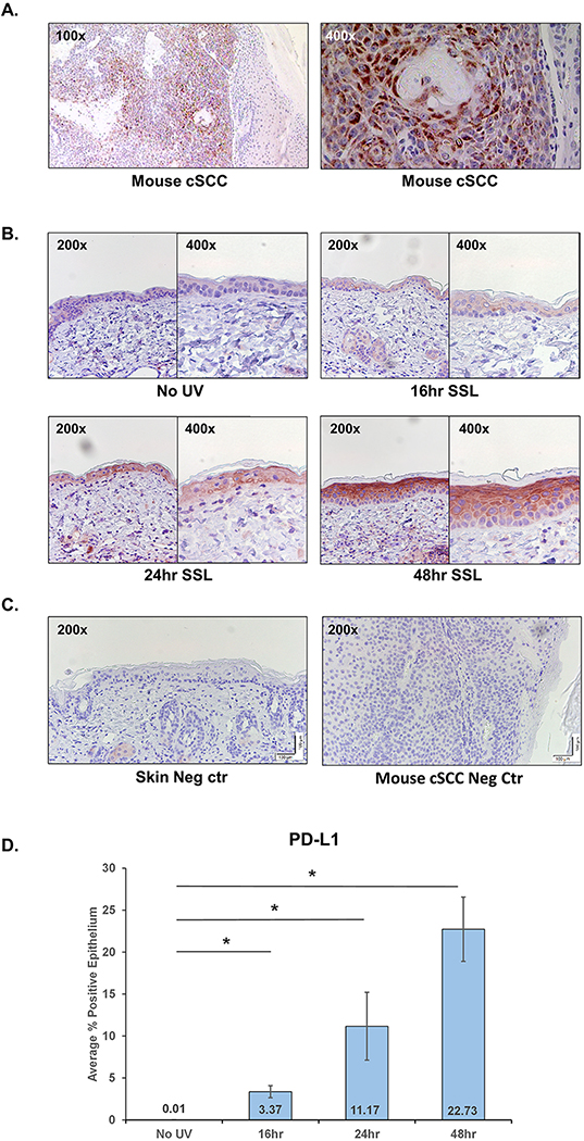
Overexpression of PD-L1 (CD274) on tumor cells may represent a hallmark of immune evasion, and overexpression has been documented in several tumors including cutaneous squamous cell carcinoma (cSCC). While PD-L1/PD-1 activity in the skin has been primarily described in inflammatory models, our goal was to examine PD-L1 expression in human keratinocytes exposed to UV irradiation. We assessed PD-L1 expression in human sun-protected (SP) and sun-damaged (SD) skin, actinic keratosis (AK), and cSCC using IHC and protein microarray. Both methods found low baseline levels of PD-L1 in SP and SD skin and significantly increased expression in cSCC. Next, we examined PD-L1 expression in acute models of UV exposure. In human SP skin exposed to 2-3 MED of UV (n = 20), epidermal PD-L1 was induced in 70% of subjects after 24 h (P = 0.0001). SKH-1 mice exposed to acute UV also showed significant epidermal PD-L1 induction at 16, 24 and 48 h. A time- and dose-dependent induction of PD-L1 was confirmed in cultured human keratinocytes after UV, which was markedly reduced in the presence of MEK/ERK, JNK or STAT3 inhibitors. These findings suggest that UV induces upregulation of PD-L1 through established, pharmacologically targetable stress-signaling pathways in keratinocytes.
© 2021 American Society for Photobiology.
Conflict of interest statement
Conflict of Interest Disclosure
The authors have no conflicts of interest to disclose.
Figures


Similar articles
-
Expression of PD-L1 in keratoacanthoma and different stages of progression in cutaneous squamous cell carcinoma.Cancer Immunol Immunother. 2017 Sep;66(9):1199-1204. doi: 10.1007/s00262-017-2015-x. Epub 2017 May 13. Cancer Immunol Immunother. 2017. PMID: 28501937 Free PMC article.
-
Inhibition of UV-Induced Stress Signaling and Inflammatory Responses in SKH-1 Mouse Skin by Topical Small-Molecule PD-L1 Blockade.JID Innov. 2024 Jan 5;4(2):100255. doi: 10.1016/j.xjidi.2023.100255. eCollection 2024 Mar. JID Innov. 2024. PMID: 38328594 Free PMC article.
-
New perspectives on role of tumor microenvironment in progression of cutaneous squamous cell carcinoma.Cell Tissue Res. 2016 Sep;365(3):691-702. doi: 10.1007/s00441-016-2457-z. Epub 2016 Jul 14. Cell Tissue Res. 2016. PMID: 27411692 Review.
-
[ASSOCIATION OF SKIN PHOTOTYPE AND UV EXPOSURE WITH EXPRESSION OF HER RECEPTORS, Ki67 AND p53 IN PATIENTS WITH CUTANEOUS SQUAMOUS CELL CARCINOMA].Acta Med Croatica. 2015;69(5):431-8. Acta Med Croatica. 2015. PMID: 29087088 Croatian.
-
TGFβ Signaling in Photoaging and UV-Induced Skin Cancer.J Invest Dermatol. 2021 Apr;141(4S):1104-1110. doi: 10.1016/j.jid.2020.11.007. Epub 2021 Jan 7. J Invest Dermatol. 2021. PMID: 33358021 Free PMC article. Review.
Cited by
-
Review: PD-L1 as an emerging target in the treatment and prevention of keratinocytic skin cancer.Mol Carcinog. 2023 Jan;62(1):52-61. doi: 10.1002/mc.23464. Epub 2022 Sep 19. Mol Carcinog. 2023. PMID: 36121318 Free PMC article. Review.
-
Association of PD-L1 Expression with Clinicopathologic Characters in Gastric Cancer: A Comprehensive Meta-analysis.Curr Med Chem. 2024;31(21):3198-3216. doi: 10.2174/0109298673263784230922060257. Curr Med Chem. 2024. PMID: 37921182
-
Photoaging: UV radiation-induced cGAS-STING signaling promotes the aging process in skin by remodeling the immune network.Biogerontology. 2025 Jun 20;26(4):123. doi: 10.1007/s10522-025-10268-1. Biogerontology. 2025. PMID: 40542276 Free PMC article. Review.
-
Optimizing the quality of horticultural crop: insights into pre-harvest practices in controlled environment agriculture.Front Plant Sci. 2024 Jul 23;15:1427471. doi: 10.3389/fpls.2024.1427471. eCollection 2024. Front Plant Sci. 2024. PMID: 39109059 Free PMC article. Review.
-
The role of the immunosuppressive PD-1/PD-L1 checkpoint pathway in the aging process and age-related diseases.J Mol Med (Berl). 2024 Jun;102(6):733-750. doi: 10.1007/s00109-024-02444-6. Epub 2024 Apr 11. J Mol Med (Berl). 2024. PMID: 38600305 Free PMC article. Review.
References
-
- Agata Y, Kawasaki A, Nishimura H, Ishida Y, Tsubata T, Yagita H, et al. Expression of the PD-1 antigen on the surface of stimulated mouse T and B lymphocytes. Int Immunol. 1996;8(5):765–72. - PubMed
-
- Franklin C, Livingstone E, Roesch A, Schilling B, Schadendorf D. Immunotherapy in melanoma: Recent advances and future directions. Eur J Surg Oncol. 2017;43(3):604–11. - PubMed
Publication types
MeSH terms
Substances
Grants and funding
LinkOut - more resources
Full Text Sources
Other Literature Sources
Research Materials
Miscellaneous
