. 2021 Jul;86(1):293-307.
doi: 10.1002/mrm.28726. Epub 2021 Feb 21.

Spatial dependency and the role of local susceptibility for velocity selective arterial spin labeling (VS-ASL) relative tagging efficiency using accelerated 3D radial sampling with a BIR-8 preparation

Affiliations

Spatial dependency and the role of local susceptibility for velocity selective arterial spin labeling (VS-ASL) relative tagging efficiency using accelerated 3D radial sampling with a BIR-8 preparation

James H Holmes et al. Magn Reson Med. 2021 Jul.

Abstract

Purpose: Velocity selective arterial spin labeling (VS-ASL) is a promising approach for non-contrast perfusion imaging that provides robustness to vascular geometry and transit times; however, VS-ASL assumes spatially uniform tagging efficiency. This work presents a mapping approach to investigate VS-ASL relative tagging efficiency including the impact of local susceptibility effects on a BIR-8 preparation.

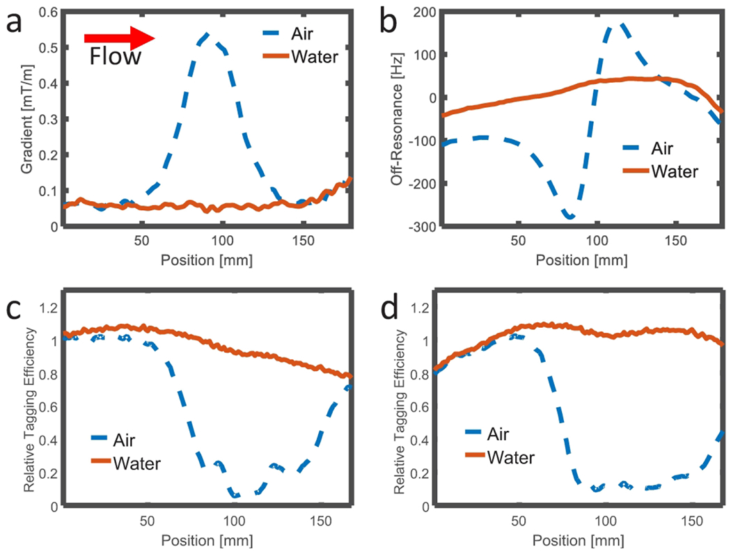
Methods: Numerical simulations of tagging efficiency were performed to evaluate sensitivity to regionally varying local susceptibility gradients and blood velocity. Tagging efficiency mapping was performed in susceptibility phantoms and healthy human subjects (N = 7) using a VS-ASL preparation module followed by a short, high spatial resolution 3D radial-based image acquisition. Tagging efficiency maps were compared to 4D-flow, B1 , and B0 maps acquired in the same imaging session for six of the seven subjects.

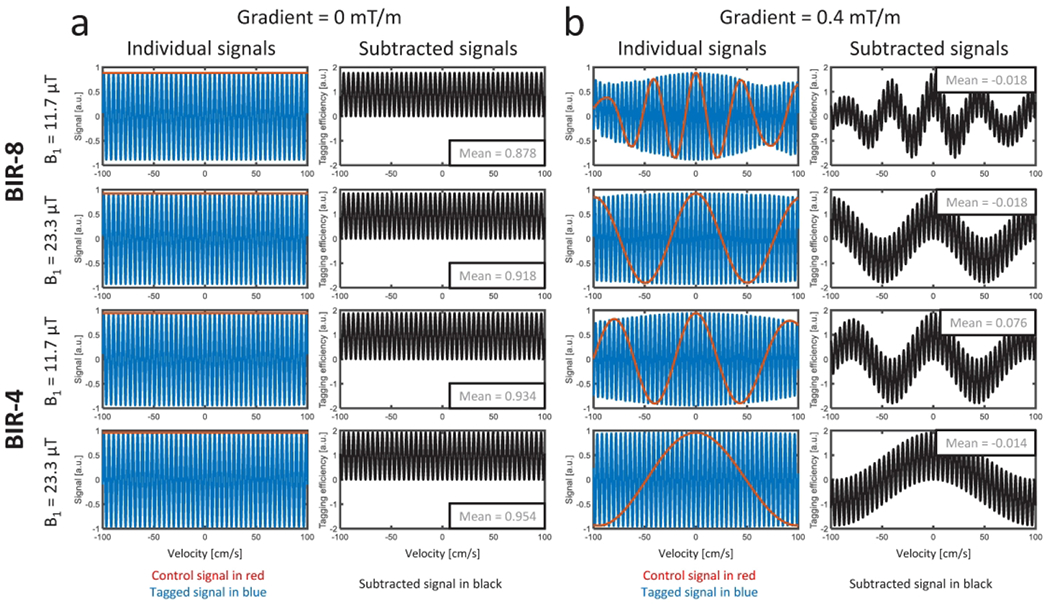
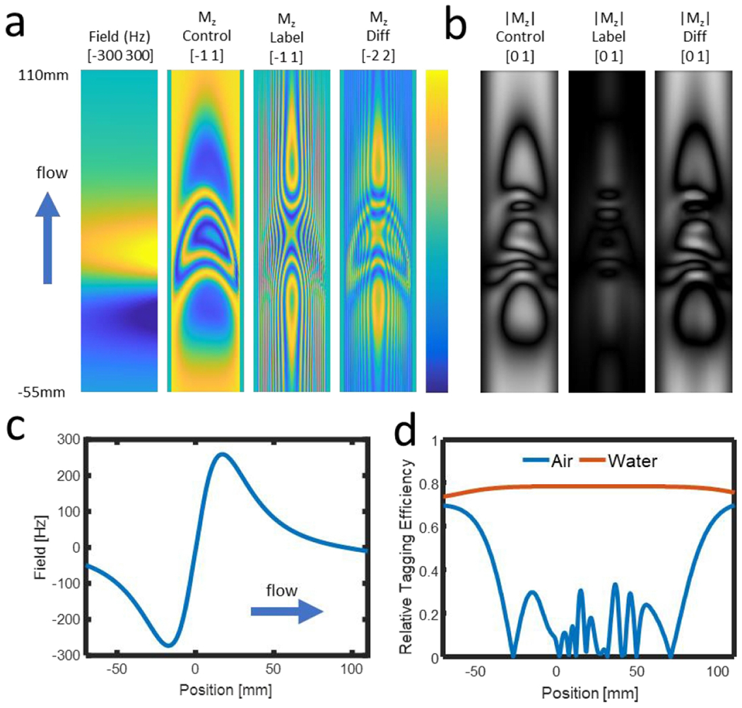
Results: Numerical simulations were found to predict reduced tagging efficiency with the combination of high blood velocity and local gradient fields. Phantom experiments corroborated numerical results. Relative efficiency mapping in normal volunteers showed unique efficiency patterns depending on individual subject anatomy and physiology. Uniform tagging efficiency was generally observed in vivo, but reduced efficiency was noted in regions of high blood velocity and local susceptibility gradients.

Conclusion: We demonstrate an approach to map the relative tagging efficiency and show application of this methodology to a novel BIR-8 preparation recently proposed in the literature. We present results showing rapid flow in the presence of local susceptibility gradients can lead to complicated signal modulations in both tag and control images and reduced tagging efficiency.

Keywords: arterial spin labeling; cerebral blood flow; perfusion; velocity-selective ASL.


Figures

Figure 1.
Figure 1.
Examples from simulations of BIR-8 and BIR-4 preparations with peak B1 of 11.7 μT and 23.3 μT, assuming a parabolic flow with 100 cm/s peak velocity, located in local gradient fields of 0 mT/m (a) and 0.4 mT/m (b). Control (red), tag (blue), and subtracted (black) signals are plotted for each. When the vessel is located in a local gradient field of 0 mT/m (a), control signal (red) is constant across all velocities. When a local gradient field is present (= 0.4 mT/m; b), perturbations are apparent in control, tag, and subtracted signals, resulting in a complex and decreased subtraction signal in parabolic flow. A more complicated pattern was observed in the complex difference signals for the longer BIR-8 preparation module with B1 peak of 11.7 μT.
Figure 2.
Figure 2.
Mean tagging efficiency predicted for parabolic flow as a function of the dot product of local susceptibility gradient and maximum velocity. This was calculated for a peak velocity of 100 cm/s but was similar for lower velocities. Note the efficiency falls off rapidly for faster flowing spins in the presence of local gradient. The reduced efficiency at higher products of velocity and susceptibility gradient was greater for the BIR-8 preparation and at lower peak B1.
Figure 3.
Figure 3.
Simulated field, and Mz images along the length of the vessel (a), with brackets providing the range of lower and upper limits of the color visualization. The control image deviates substantially from the ideal uniform value of 1. When considering intravoxel signal averaging at 1 mm resolution (b) this modulation results in signal loss in the control image that results in lower signal difference despite the effective signal saturation in the label image. Taking the field values along the centerline of the vessel (c) and the resulting signal at 1 mm resolution with air and water filled wells (d), substantial signal loss is observed that is shifted downstream from the field perturbation. These results are in good agreement with those observed in physical phantoms (Figure 4).
Figure 4.
Figure 4.
Geometry used for the physical phantom that included a susceptibility well (far left). Control, tag, and magnetic field gradient images from an agar/water gel phantom with flow at 500 mL/min, adjacent to a susceptibility insert made up of wells of either water (left) or air (right). Signal in the tube remains low for both susceptibility wells; however, the control image shows signal loss near the air susceptibility well. Correspondingly, the magnetic field gradient is much greater near the air susceptibility well whereas there is no significant gradient due to the similar susceptibility of the agar/water phantom and the water filled well.
Figure 5.
Figure 5.
Profiles of the absolute value of the magnetic field gradient (a) and off-resonance frequency (b) measured along the centerline in the direction of flow through the tube located adjacent to the air (blue line) and water (red line) susceptibility phantoms. Corresponding profiles of the relative tagging efficiency are plotted for flow rates of 500 mL/min (c) and 1000 mL/min (d). Greater local gradient is measured along the tube adjacent to the air susceptibility well and the relative tagging efficiency is much lower in this region and immediately downstream (blue lines).
Figure 6.
Figure 6.
Limited MIPs from the relative tagging efficiency maps for each volunteer including coronal (top row) and sagittal (bottom row) reformats. Similar patterns of high relative tagging efficiency are observed in the sagittal sinus, lower ICAs and BA; however, reduced relative efficiency is observed adjacent to the nasal sinus including the terminus (white arrows) and middle (yellow arrows) ICAs. Note that signal from structures including vessel segments located outside of the limited volume used for the MIP projection are not displayed.
Figure 7.
Figure 7.
Axial and images from two different sagittal slice locations from a typical volunteer as well as limited thickness MIPs. Signal loss is visible in the middle cerebral artery (MCAs) (arrow) of the control image. The axial and sagittal difference images show high subtracted signal in the blood vessels except for in the MCAs (arrow). VS-ASL relative tagging efficiency maps show high efficiency in both the arterial and venous systems; however, the arteries over the cavernous sinus are not well tagged (arrow).
Figure 8.
Figure 8.
Box plots showing regional measurements for a) B0, b) B1, c) Gradient dot product, d) magnitude of velocity, e) magnitude of velocity in z-direction, and f) relative tagging efficiency. Regions were derived from segments defined in Supporting Information Figure S3.
Figure 9.
Figure 9.
Sagittal gradient field maps (a) and MIP angiograms (b) acquired with 3 external shim gradient settings in the S/I direction (0.00 mT/m, 0.08 mT/m, and 0.17 mT/m from left to right). Signal dropout is seen in the region of rapid flow over the nasal sinus for all shim gradient values (b, arrows) with greater lost in other fast flowing vessels with increasing local gradient.

References

    1. van Osch MJP, van der Grond J, Bakker CJG. Partial volume effects on arterial input functions: shape and amplitude distortions and their correction. J Magn Reson Imaging. 2005;22(6):704–709. doi:10.1002/jmri.20455 - DOI - PubMed
    1. Jochimsen TH, Newbould RD, Skare ST, et al. Identifying systematic errors in quantitative dynamic-susceptibility contrast perfusion imaging by high-resolution multi-echo parallel EPI. NMR in Biomedicine. 2007;20(4):429–438. doi:10.1002/nbm.1107 - DOI - PMC - PubMed
    1. Alsop DC, Detre JA. Reduced transit-time sensitivity in noninvasive magnetic resonance imaging of human cerebral blood flow. J Cereb Blood Flow Metab. 1996;16(6):1236–1249. doi:10.1097/00004647-199611000-00019 - DOI - PubMed
    1. Deibler AR, Pollock JM, Kraft RA, Tan H, Burdette JH, Maldjian JA. Arterial spin-labeling in routine clinical practice, part 1: technique and artifacts. AJNR Am J Neuroradiol. 2008;29(7):1228–1234. doi:10.3174/ajnr.A1030 - DOI - PMC - PubMed
    1. Zaharchuk G, Bammer R, Straka M, et al. Arterial spin-label imaging in patients with normal bolus perfusion-weighted MR imaging findings: pilot identification of the borderzone sign. Radiology. 2009;252(3):797–807. doi:10.1148/radiol.2523082018 - DOI - PMC - PubMed

Publication types

LinkOut - more resources