

Biplanar Videoradiography to Study the Wrist and Distal Radioulnar Joints
- PMID: 33616093
- PMCID: PMC8182367
- DOI: 10.3791/62102
Biplanar Videoradiography to Study the Wrist and Distal Radioulnar Joints
Abstract
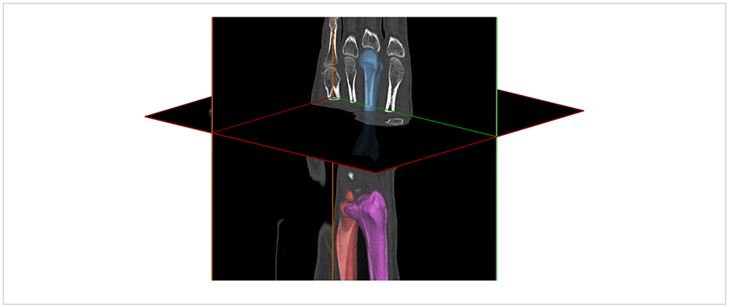
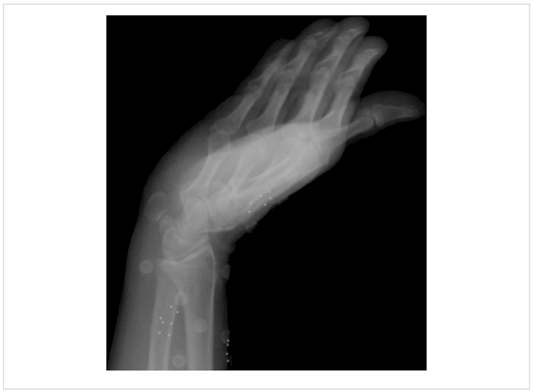
Accurate measurement of skeletal kinematics in vivo is essential for understanding normal joint function, the influence of pathology, disease progression, and the effects of treatments. Measurement systems that use skin surface markers to infer skeletal motion have provided important insight into normal and pathological kinematics, however, accurate arthrokinematics cannot be attained using these systems, especially during dynamic activities. In the past two decades, biplanar videoradiography (BVR) systems have enabled many researchers to directly study the skeletal kinematics of the joints during activities of daily living. To implement BVR systems for the distal upper extremity, videoradiographs of the distal radius and the hand are acquired from two calibrated X-ray sources while a subject performs a designated task. Three-dimensional (3D) rigid-body positions are computed from the videoradiographs via a best-fit registrations of 3D model projections onto to each BVR view. The 3D models are density-based image volumes of the specific bone derived from independently acquired computed-tomography data. Utilizing graphics processor units and high-performance computing systems, this model-based tracking approach is shown to be fast and accurate in evaluating the wrist and distal radioulnar joint biomechanics. In this study, we first summarized the previous studies that have established the submillimeter and subdegree agreement of BVR with an in vitro optical motion capture system in evaluating the wrist and distal radioulnar joint kinematics. Furthermore, we used BVR to compute the center of rotation behavior of the wrist joint, to evaluate the articulation pattern of the components of the implant upon one another, and to assess the dynamic change of ulnar variance during pronosupination of the forearm. In the future, carpal bones may be captured in greater detail with the addition of flat panel X-ray detectors, more X-ray sources (i.e., multiplanar videoradiography), or advanced computer vision algorithms.
Conflict of interest statement
Disclosures
We have no conflict of interest to declare.
Figures


References
-
- Leardini A, Chiari L, Croce UD, Cappozzo A Human movement analysis using stereophotogrammetry: Part 3. Soft tissue artifact assessment and compensation. Gait & Posture. 21 (2), 212–225 (2005). - PubMed
-
- Tashman S, Anderst W In vivo measurement of dynamic joint motion using high speed biplane radiography and CT: application to canine ACL deficiency. Journal of Biomechanical Engineering. 125 (2), 238–245 (2003). - PubMed
-
- Moore DC et al. Computed Tomography Image-Based Kinematic Analysis: An Overview. Handbook of Imaging in Biological Mechanics. 115–126 (2014).
Publication types
MeSH terms
Grants and funding
LinkOut - more resources
Full Text Sources
Other Literature Sources
Medical
Miscellaneous
