

Hedgehog Signaling Pathway Regulates the Proliferation and Differentiation of Rat Meibomian Gland Epithelial Cells
- PMID: 33616621
- PMCID: PMC7910630
- DOI: 10.1167/iovs.62.2.33
Hedgehog Signaling Pathway Regulates the Proliferation and Differentiation of Rat Meibomian Gland Epithelial Cells
Abstract
Purpose: Meibomian glands play a vital role in maintaining ocular surface stability. This study aimed to investigate whether Hedgehog signaling is involved in the regulation of meibomian gland epithelial cells.
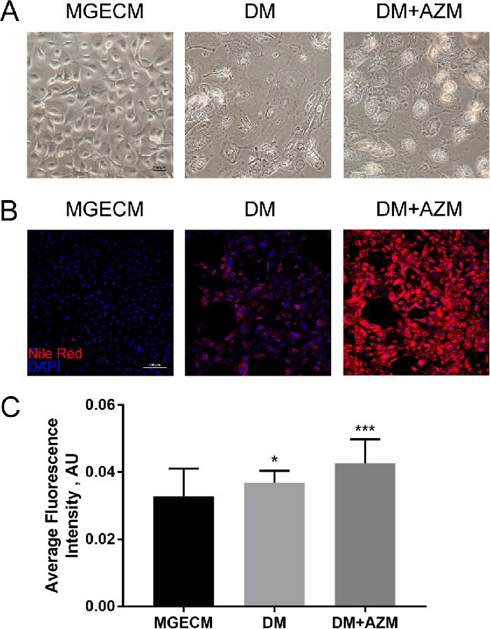
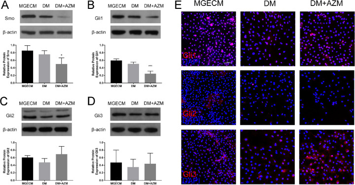
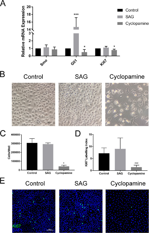
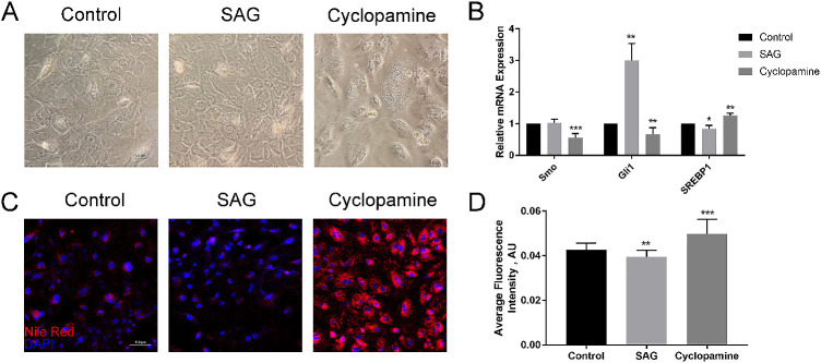
Methods: Rat meibomian glands epithelial cells (RMGECs) were isolated from ducts and ductules, and then were cultivated to passage two on Matrigel coated wells in meibomian gland epithelial cells medium (MGECM). Cells were switched from MGECM to differentiation medium (DM) or DM added 10 µg/mL azithromycin (DM + AZM) when reached 50% to 60% confluence. The effects of the Smoothened (Smo) agonist (Smo agonist [SAG]) and antagonist (by cyclopamine) on RMGECs were analyzed using quantitative RT-PCR, cell proliferation analysis, immunofluorescence staining, and Nile red staining.
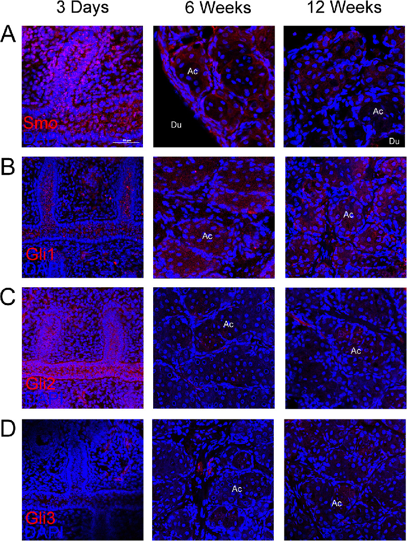
Results: The Hedgehog receptor, Smo, and its downstream molecules, Glis, were expressed both in vivo and in vitro. Smo and Gli1 both decreased with the increase of differentiation in vitro. Smo antagonist, cyclopamine, reduced cell numbers, and the expression of Ki67 in MGECM, and promoted the expression of SREBP1 and lipid production in DM + AZM. Smo agonist, SAG, inhibited the expression of SREBP1 and lipid accumulation in DM + AZM but showed no significant effects on raising cell numbers and the expression of Ki67 in MGECM.
Conclusions: The Hedgehog signaling pathway appears to play important roles in RMGECs proliferation and differentiation. This may provide a potential therapeutic way to treat meibomian gland dysfunction (MGD).
Conflict of interest statement
Disclosure:
Figures
Similar articles
-
Organotypic Culture of Mouse Meibomian Gland: A Novel Model to Study Meibomian Gland Dysfunction In Vitro.Invest Ophthalmol Vis Sci. 2020 Apr 9;61(4):30. doi: 10.1167/iovs.61.4.30. Invest Ophthalmol Vis Sci. 2020. PMID: 32330227 Free PMC article.
-
Er-Dong-Xiao-Ke decoction regulates lipid metabolism via PPARG-mediated UCP2/AMPK signaling to alleviate diabetic meibomian gland dysfunction.J Ethnopharmacol. 2024 Oct 28;333:118484. doi: 10.1016/j.jep.2024.118484. Epub 2024 Jun 24. J Ethnopharmacol. 2024. PMID: 38925318
-
The inhibition of p38 MAPK blocked inflammation to restore the functions of rat meibomian gland epithelial cells.Exp Eye Res. 2023 Jun;231:109470. doi: 10.1016/j.exer.2023.109470. Epub 2023 Apr 13. Exp Eye Res. 2023. PMID: 37059216
-
Human meibomian gland epithelial cell culture models: Current progress, challenges, and future directions.Ocul Surf. 2022 Jan;23:96-113. doi: 10.1016/j.jtos.2021.11.012. Epub 2021 Nov 26. Ocul Surf. 2022. PMID: 34843998 Review.
-
In vivo Meibomian gland imaging techniques: A review of the literature.J Fr Ophtalmol. 2020 Apr;43(4):e123-e131. doi: 10.1016/j.jfo.2019.11.003. Epub 2020 Jan 9. J Fr Ophtalmol. 2020. PMID: 31928786 Review.
Cited by
-
Catalpol inhibits Hedgehog signaling pathway to suppress proliferation and promote lipid accumulation in rat meibomian gland epithelial cells.Cytotechnology. 2025 Jun;77(3):105. doi: 10.1007/s10616-025-00769-9. Epub 2025 May 20. Cytotechnology. 2025. PMID: 40406032
-
Identification of Meibomian gland stem cell populations and mechanisms of aging.Nat Commun. 2025 Feb 15;16(1):1663. doi: 10.1038/s41467-025-56907-6. Nat Commun. 2025. PMID: 39955307 Free PMC article.
-
Isolation and Culture of Human Meibomian Gland Ductal Cells.Invest Ophthalmol Vis Sci. 2023 Dec 1;64(15):29. doi: 10.1167/iovs.64.15.29. Invest Ophthalmol Vis Sci. 2023. PMID: 38133507 Free PMC article.
-
Identification of Meibomian gland stem cell populations and mechanisms of aging.bioRxiv [Preprint]. 2024 Aug 10:2024.08.09.607015. doi: 10.1101/2024.08.09.607015. bioRxiv. 2024. Update in: Nat Commun. 2025 Feb 15;16(1):1663. doi: 10.1038/s41467-025-56907-6. PMID: 39149265 Free PMC article. Updated. Preprint.
-
Single-Cell Ribonucleic Acid Sequencing Clarifies Cold Tolerance Mechanisms in the Pacific White Shrimp (Litopenaeus Vannamei).Front Genet. 2022 Jan 12;12:792172. doi: 10.3389/fgene.2021.792172. eCollection 2021. Front Genet. 2022. PMID: 35096009 Free PMC article.
References
-
- Asano N, Hampel U, Garreis F, et al. .. Differentiation patterns of immortalized human meibomian gland epithelial cells in three-dimensional culture. Invest Ophthalmol Vis Sci. 2018; 59: 1343–1353. - PubMed
Publication types
MeSH terms
Substances
LinkOut - more resources
Full Text Sources
Other Literature Sources
Miscellaneous
