. 2021 Jan;109(1):99-114.
doi: 10.1002/JLB.5COVA0820-520RRR.

Flow cytometric evaluation of the neutrophil compartment in COVID-19 at hospital presentation: A normal response to an abnormal situation

Affiliations

Flow cytometric evaluation of the neutrophil compartment in COVID-19 at hospital presentation: A normal response to an abnormal situation

Roy Spijkerman et al. J Leukoc Biol. 2021 Jan.

Abstract

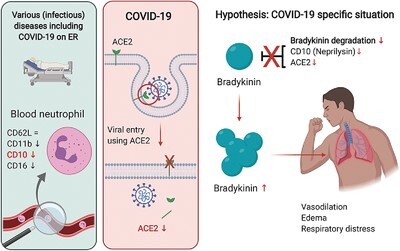
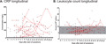
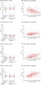
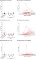
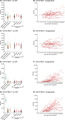
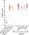
Coronavirus disease 2019 (COVID-19) is a rapidly emerging pandemic disease caused by the severe acute respiratory syndrome coronavirus 2 (SARS-CoV-2). Critical COVID-19 is thought to be associated with a hyper-inflammatory process that can develop into acute respiratory distress syndrome, a critical disease normally mediated by dysfunctional neutrophils. This study tested the hypothesis whether the neutrophil compartment displays characteristics of hyperinflammation in COVID-19 patients. Therefore, a prospective study was performed on all patients with suspected COVID-19 presenting at the emergency room of a large academic hospital. Blood drawn within 2 d after hospital presentation was analyzed by point-of-care automated flow cytometry and compared with blood samples collected at later time points. COVID-19 patients did not exhibit neutrophilia or eosinopenia. Unexpectedly neutrophil activation markers (CD11b, CD16, CD10, and CD62L) did not differ between COVID-19-positive patients and COVID-19-negative patients diagnosed with other bacterial/viral infections, or between COVID-19 severity groups. In all patients, a decrease was found in the neutrophil maturation markers indicating an inflammation-induced left shift of the neutrophil compartment. In COVID-19 this was associated with disease severity.

Keywords: CD10; SARS‐CoV‐2; activation; flow cytometry; neprilysin; neutrophil.


Figures

None
Graphical abstract
FIGURE 1
FIGURE 1
Approach for flow cytometric analysis. (A) Fluorescence minus one (FMO) controls of the used panel of markers (CD16, CD11b, CD62L, CD10). CD64 was also tested in this panel but is not shown here because it is not discussed further in this article; (B) gating strategy for analyzing the blood samples from healthy controls and patients. Flow cytometric analysis was done using FlowJo analysis software (Tree Star Inc.)
FIGURE 2
FIGURE 2
Flow diagram depicting inclusion and exclusion of patients in this study
FIGURE 3
FIGURE 3
Absolute cell counts of white blood cell populations in the ER. Absolute cell counts of (A) Total white blood cells; (B) Neutrophils; (C) Lymphocytes; (D); Monocytes and (E) Eosinophils in peripheral blood of healthy controls (HC, n = 23), patients with other diagnoses categorized as bacterial (n = 84) or viral (n = 17) and coronavirus disease 2019 (COVID-19) patients as a total group (n = 103) and categorized according to the WHO classification system for COVID-19 as moderate (n = 10), severe (n = 16), or critical (n = 77) disease. The normal range of cell counts is displayed by the gray area. Results of the statistical analysis are shown in Supporting Information Table 1.1. Data are represented as individual dots with medians and interquartile range (IQR)
FIGURE 4
FIGURE 4
Plasma concentrations of (A) C-reactive protein (CRP) and (B) leukocytes over time after the onset of symptoms in coronavirus disease 2019 (COVID-19) patients (n = 78) with critical or moderate disease. The normal range of cell counts is displayed by the gray area. Data are represented as individual black dots. If multiple measurements were done for one patient, different measurements are connected with a red line to indicate paired data. Data were retrieved from the patients’ records
FIGURE 5
FIGURE 5
Neutrophil activation markers CD62L and CD11b in neutrophils in the absence and presence of a bacterial stimulus (N-Formyl-norleucyl-leucyl-phenylalanine [fNLF], 10 μM) to induce activation, as measured in the ER and over time (longitudinal). Flow cytometric measurements were done in the ER using peripheral blood from patients with other diagnoses categorized as bacterial (n = 84) or viral (n = 17) infections and from coronavirus disease 2019 (COVID-19) patients as a total group (n = 103) and categorized according to the WHO classification system for COVID-19 as moderate (n = 10), severe (n = 16), or critical (n = 77) disease. A group of healthy controls (n = 23) was also included. Longitudinal measurements were done with peripheral blood from patients on the COVID-19 ward (n = 78) who were categorized as having moderate or critical disease. For the majority of patients multiple measurements were done overtime. (A) CD62L without fNLF in the ER; (B) CD62L without fNLF longitudinal; (C) CD62L with fNLF in the ER; (D) CD62L with fNLF longitudinal; (E) CD11b without fNLF in the ER; (F) CD11b without fNLF longitudinal; (G) CD11b with fNLF in the ER; and (H) CD11b with fNLF longitudinal. Results of the statistical analysis are shown in Supporting Information Table S1.2. Data are represented as individual dots with medians and interquartile range (IQR; ER samples) or as individual data points connected with a red line to indicate paired data (longitudinal data)
FIGURE 6
FIGURE 6
Absolute cell counts of neutrophil progenitors gated based on CD11b/CD16 expression as measured in the ER and over time (longitudinal). Flow cytometric measurements were done in the ER using peripheral blood from patients with other diagnoses categorized as bacterial (n = 84) or viral (n = 17) infections and coronavirus disease 2019 (COVID-19) patients as a total group (n = 103) and categorized according to the WHO classification system for COVID-19 as moderate (n = 10), severe (n = 16), or critical (n = 77) disease. A group of healthy controls (n = 23) was also included. Longitudinal measurements were done with peripheral blood from patients on the COVID-19 ward (n = 78) who were categorized as having moderate or critical disease. For the majority of patients multiple measurements were done overtime. (A) Promyelocyte counts in the ER; (B) promyelocyte counts longitudinal; (C) myelocyte counts on the ER; (D) myelocyte counts longitudinal; (E) metamyelocyte counts on the ER; and (F) metamyelocyte counts longitudinal. Results of the statistical analysis are shown in Supporting Information Table S1.3. Data are represented as individual dots with medians and interquartile range (IQR; ER samples) or as individual data points connected with a red line to indicate paired data (longitudinal data)
FIGURE 7
FIGURE 7
Expression of neutrophil maturation markers CD16 and CD10 in the absence and presence of a bacterial stimulus (N-Formyl-norleucyl-leucyl-phenylalanine [fNLF], 10 μM) to induce activation, as measured in the ER and over time (longitudinal). Flow cytometric measurements were done in the ER using peripheral blood from patients with other diagnoses categorized as bacterial (n = 84) or viral (n = 17) infections and coronavirus disease 2019 (COVID-19) patients as a total group (n = 103) and categorized according to the WHO classification system for COVID-19 as moderate (n = 10), severe (n = 16), or critical (n = 77) disease. A group of healthy controls (n = 23) was also included. Longitudinal measurements were done with peripheral blood from patients on the COVID-19 ward (n = 78) who were categorized as having moderate or critical disease. For the majority of patients multiple measurements were done overtime. (A) CD16 without fNLF in the ER; (B) CD16 without fNLF longitudinal; (C) CD16 with fNLF in the ER; (D) CD16 with fNLF longitudinal; (E) CD10 without fNLF in the ER; (F) CD10 without fNLF longitudinal; (G) CD10 with fNLF in the ER; and (H) CD10 with fNLF longitudinal. Results of the statistical analysis are shown in Supporting Information Table S1.4. Data are represented as individual dots with medians and interquartile range (IQR; ER samples) or as individual data points connected with a red line to indicate paired data (longitudinal data)
FIGURE 8
FIGURE 8
Multidimensional analysis of neutrophil characteristics in coronavirus disease 2019 (COVID-19) patients compared to controls. Based on the differences in neutrophil characteristics in the presence and absence of N-Formyl-norleucyl-leucyl-phenylalanine (fNLF; 10 μM) present in COVID-19 patients compared to controls, a discriminant analysis of multi-aspect cytometry (DAMACY) score is calculated. A higher score indicated more deviations in neutrophil marker expression compared to healthy controls. DAMACY scores are shown for healthy controls (n = 23), patients with other diagnoses categorized as bacterial (n = 84) or viral (n = 17) and COVID-19 patients as a total group (n = 103) and categorized according to the WHO classification system for COVID-19 as moderate (n = 10), severe (n = 16), or critical (n = 77) disease

References

    1. Hua J, Shaw R. Corona virus (COVID-19) "infodemic" and emerging issues through a data lens: the case of China. Int J Environ Res Public Health;17. 10.3390/ijerph17072309. Epub ahead of print 2020. - DOI - PMC - PubMed
    1. Jordan RE, Adab P, Cheng KK. Covid-19: risk factors for severe disease and death. BMJ. 2020;368:m1198. 10.1136/bmj.m1198. - DOI - PubMed
    1. Guan W, Ni Z, Hu Y, et al. Clinical characteristics of coronavirus disease 2019 in China. N Engl J Med. 10.1056/NEJMoa2002032. Epub ahead of print 2020. - DOI - PMC - PubMed
    1. Wang Y, Wang Y, Chen Y, et al. Unique epidemiological and clinical features of the emerging 2019 novel coronavirus pneumonia (COVID-19) implicate special control measures. J Med Virol. 2020;92:568-576. - PMC - PubMed
    1. Tang N, Li D, Wang X, et al. Abnormal coagulation parameters are associated with poor prognosis in patients with novel coronavirus pneumonia. J Thromb Haemost. 2020;18:844-847. - PMC - PubMed

Publication types

MeSH terms