

A Coumarin-Based Analogue of Thiacetazone as Dual Covalent Inhibitor and Potential Fluorescent Label of HadA in Mycobacterium tuberculosis
- PMID: 33617235
- PMCID: PMC8022203
- DOI: 10.1021/acsinfecdis.0c00325
A Coumarin-Based Analogue of Thiacetazone as Dual Covalent Inhibitor and Potential Fluorescent Label of HadA in Mycobacterium tuberculosis
Abstract
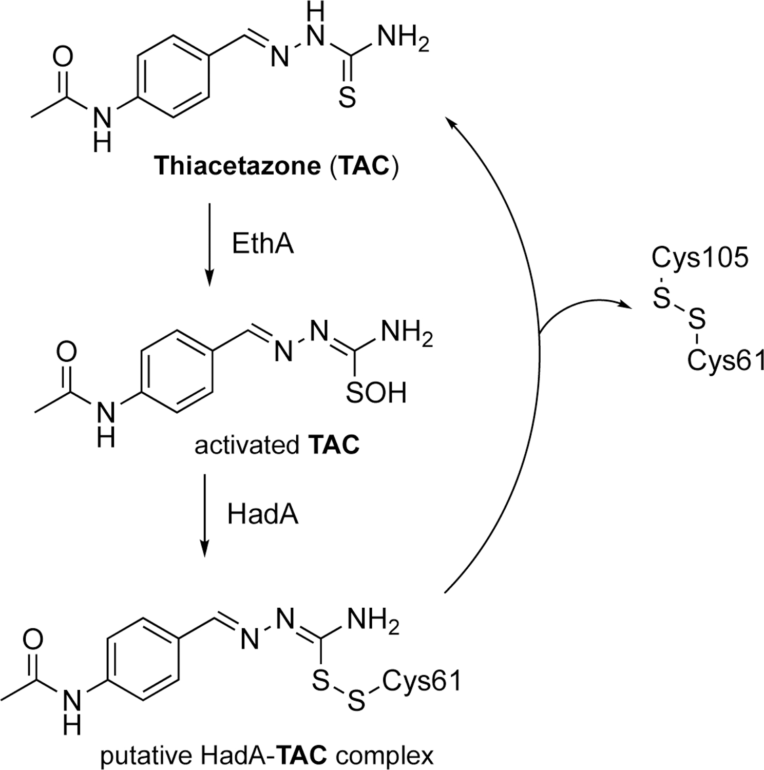
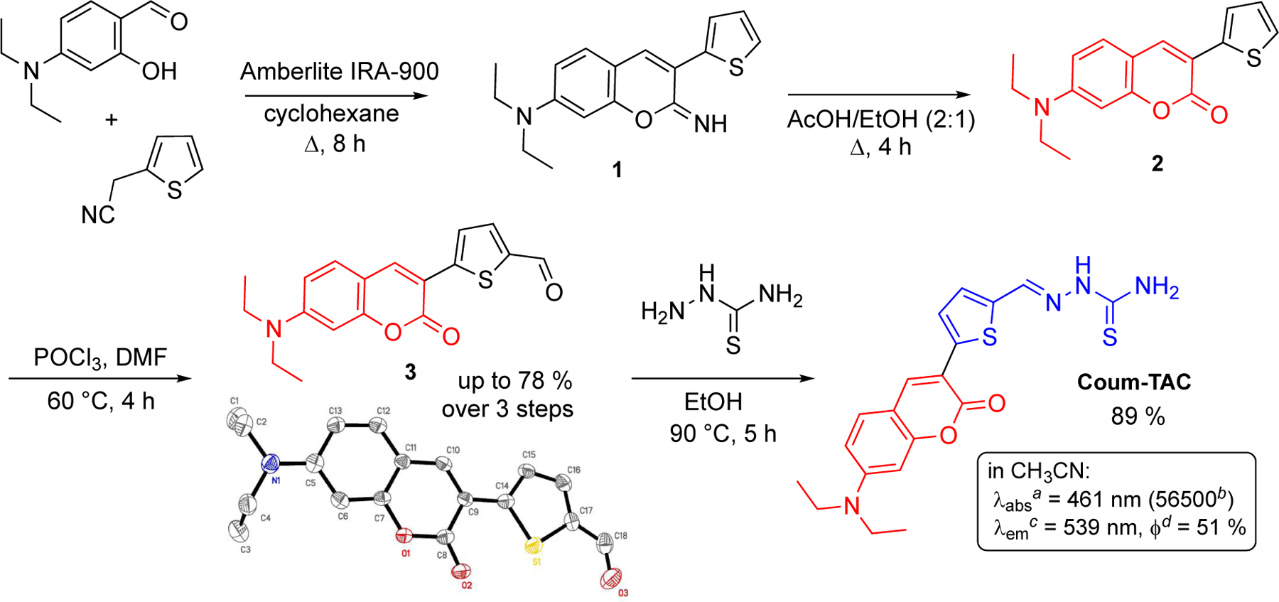
A novel coumarin-based molecule, designed as a fluorescent surrogate of a thiacetazone-derived antitubercular agent, was quickly and easily synthesized from readily available starting materials. This small molecule, coined Coum-TAC, exhibited a combination of appropriate physicochemical and biological properties, including resistance toward hydrolysis and excellent antitubercular efficiency similar to that of well-known thiacetazone derivatives, as well as efficient covalent labeling of HadA, a relevant therapeutic target to combat Mycobacterium tuberculosis. More remarkably, Coum-TAC was successfully implemented as an imaging probe that is capable of labeling Mycobacterium tuberculosis in a selective manner, with an enrichment at the level of the poles, thus giving for the first time relevant insights about the polar localization of HadA in the mycobacteria.
Keywords: HadA; Mycobacterium tuberculosis; coumarin; fluorescence; thiacetazone.
Figures


Similar articles
-
Synthesis, antitubercular activity and mechanism of resistance of highly effective thiacetazone analogues.PLoS One. 2013;8(1):e53162. doi: 10.1371/journal.pone.0053162. Epub 2013 Jan 3. PLoS One. 2013. PMID: 23301038 Free PMC article.
-
A common mechanism of inhibition of the Mycobacterium tuberculosis mycolic acid biosynthetic pathway by isoxyl and thiacetazone.J Biol Chem. 2012 Nov 9;287(46):38434-41. doi: 10.1074/jbc.M112.400994. Epub 2012 Sep 21. J Biol Chem. 2012. PMID: 23002234 Free PMC article.
-
In vitro activity of thiacetazone on mycobacterial species belonging to the Mycobacterium tuberculosis complex.Int J Tuberc Lung Dis. 2002 Oct;6(10):933-5. Int J Tuberc Lung Dis. 2002. PMID: 12365582
-
Coumarin as a Privileged and Medicinally Important Scaffold in the Treatment of Tuberculosis.Curr Top Med Chem. 2023;23(16):1489-1502. doi: 10.2174/1568026623666230330084058. Curr Top Med Chem. 2023. PMID: 37005527 Review.
-
Recent progress in the drug development of coumarin derivatives as potent antituberculosis agents.Eur J Med Chem. 2015 Jul 15;100:257-69. doi: 10.1016/j.ejmech.2015.06.017. Epub 2015 Jun 9. Eur J Med Chem. 2015. PMID: 26112067 Review.
Cited by
-
Identification of Novel Inhibitor of Enoyl-Acyl Carrier Protein Reductase (InhA) Enzyme in Mycobacterium tuberculosis from Plant-Derived Metabolites: An In Silico Study.Antibiotics (Basel). 2022 Aug 1;11(8):1038. doi: 10.3390/antibiotics11081038. Antibiotics (Basel). 2022. PMID: 36009907 Free PMC article.
-
Discovery of 2-(furan-2-ylmethylene)hydrazine-1-carbothioamide derivatives as novel inhibitors of SARS-CoV-2 main protease.Eur J Med Chem. 2022 Aug 5;238:114508. doi: 10.1016/j.ejmech.2022.114508. Epub 2022 Jun 3. Eur J Med Chem. 2022. PMID: 35688005 Free PMC article.
-
Anti-tuberculosis drug development via targeting the cell envelope of Mycobacterium tuberculosis.Front Microbiol. 2022 Dec 21;13:1056608. doi: 10.3389/fmicb.2022.1056608. eCollection 2022. Front Microbiol. 2022. PMID: 36620019 Free PMC article. Review.
-
Synthesis of Novel Artemisinin, Ciprofloxacin, and Norfloxacin Hybrids with Potent Antiplasmodial Activity.Antibiotics (Basel). 2024 Feb 1;13(2):142. doi: 10.3390/antibiotics13020142. Antibiotics (Basel). 2024. PMID: 38391528 Free PMC article.
-
Bioinformatic Mining and Structure-Activity Profiling of Baeyer-Villiger Monooxygenases from Mycobacterium tuberculosis.mSphere. 2022 Apr 27;7(2):e0048221. doi: 10.1128/msphere.00482-21. Epub 2022 Mar 17. mSphere. 2022. PMID: 35296143 Free PMC article.
References
-
- Grobusch MP and Kapata N (2018) Global burden of tuberculosis: where we are and what to do. Lancet Infect. Dis 18, 1291–1293. - PubMed
-
- Vilchèze C, Morbidoni HR, Weisbrod TR, Iwamoto H, Kuo M, Sacchettini JC, Jacobs WR (2000) Inactivation of the inhA-encoded fatty acid synthase II (FAS-II) enoyl-acyl carrier protein reductase induces accumulation of the FAS-I end products and cell lysis of Mycobacterium smegmatis. J. Bacteriol 182, 4059–4067. - PMC - PubMed
-
- Slama N, Jamet S, Frigui W, Pawlik A, Bottai D, Laval F, Constant P, Lemassu A, Cam K, Daffé M Brosch R, Eynard N, and Quémard A (2016) The changes in mycolic acid structures caused by hadC mutation have a dramatic effect on the virulence of Mycobacterium tuberculosis. Mol. microbial 99, 794–807. - PubMed
Publication types
MeSH terms
Substances
Grants and funding
LinkOut - more resources
Full Text Sources
Other Literature Sources
