

Can we use intraoperative high-frequency oscillations to guide tumor-related epilepsy surgery?
- PMID: 33617688
- PMCID: PMC8248094
- DOI: 10.1111/epi.16845
Can we use intraoperative high-frequency oscillations to guide tumor-related epilepsy surgery?
Abstract
Objective: In people with low-grade intrinsic brain tumors, an epileptic focus is often located close to the lesion. High-frequency oscillations (HFOs) in electrocorticography (ECoG) might help to delineate this focus. We investigated the relationship between HFOs and low-grade brain tumors and their potential value for tumor-related epilepsy surgery.
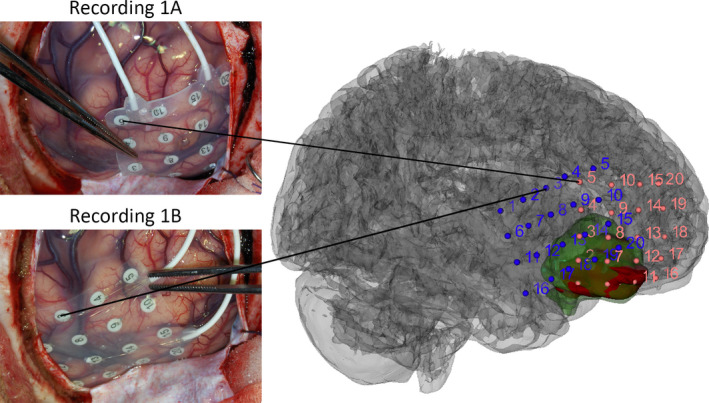
Methods: We analyzed pre- and postresection intraoperative ECoG in 41 patients with refractory epilepsy and a low-grade lesion. Electrodes were designated as overlying the tumor, adjacent resected tissue (peritumoral), or outside the resection bed using magnetic resonance imaging (MRI) and intraoperative photographs. We then used a semiautomated approach to detect HFOs as either ripples (80-250 Hz) or fast ripples (250-500 Hz).
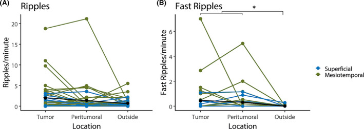
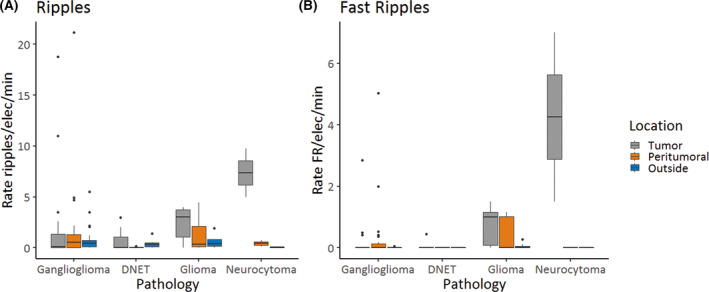
Results: The rate of fast ripples was higher in electrodes covering tumor and peritumoral tissue than outside the resection (p = .04). Mesiotemporal tumors showed more ripples (p = .002), but not more fast ripples (p = .07), than superficial tumors. Rates of fast ripples were higher in glioma and extraventricular neurocytoma than in ganglioglioma or dysembryoplastic neuroepithelial tumor (DNET). The rate of ripples and fast ripples in postresection ECoG was not higher in patients with residual tumor tissue on MRI than those without. The rate of ripples in postresection ECoG was higher in patients with good than bad seizure outcome (p = .03). Fast ripples outside the resection and in post-ECoG seem related to seizure recurrence.
Significance: Fast ripples in intraoperative ECoG can be used to help guide resection in tumor-related epilepsy surgery. Preresection fast ripples occur predominantly in epileptogenic tumor and peritumoral tissue. Fast ripple rates are higher in glioma and extraventricular neurocytoma than in ganglioglioma and DNET.
Keywords: corticography; epilepsy surgery; high-frequency oscillations; tumor-related epilepsy.
© 2021 The Authors. Epilepsia published by Wiley Periodicals LLC on behalf of International League Against Epilepsy.
Conflict of interest statement
None of the authors has any conflict of interest to disclose.
Figures
References
-
- van Breemen MSM, Wilms EB, Vecht CJ. Epilepsy in patients with brain tumours: epidemiology, mechanisms, and management. Lancet Neurol. 2007;6(5):421–30. - PubMed
-
- Pallud J, Audureau E, Blonski M, Sanai N, Bauchet L, Fontaine D, et al. Epileptic seizures in diffuse low‐grade gliomas in adults. Brain. 2014;137(2):449–62. - PubMed
-
- Chaichana KL, Parker SL, Olivi A, Quiñones‐Hinojosa A. Long‐term seizure outcomes in adult patients undergoing primary resection of malignant brain astrocytomas: clinical article. J Neurosurg. 2009;111(2):282–92. - PubMed
-
- Chang EF, Potts MB, Keles GE, Lamborn KR, Chang SM, Barbaro NM, et al. Seizure characteristics and control following resection in 332 patients with low‐grade gliomas. J Neurosurg. 2008;108(2):227–35. - PubMed
-
- Minkin K, Klein O, Mancini J, Lena G. Surgical strategies and seizure control in pediatric patients with dysembryoplastic neuroepithelial tumors: a single‐institution experience. J Neurosurg Pediatr. 2008;1(3):206–10. - PubMed
Publication types
MeSH terms
Grants and funding
LinkOut - more resources
Full Text Sources
Other Literature Sources
Medical
Research Materials
