. 2021 Mar 19;433(6):166881.
doi: 10.1016/j.jmb.2021.166881. Epub 2021 Feb 20.

Binding Dynamics of Disordered Linker Histone H1 with a Nucleosomal Particle

Affiliations

Binding Dynamics of Disordered Linker Histone H1 with a Nucleosomal Particle

Hao Wu et al. J Mol Biol. .

Abstract

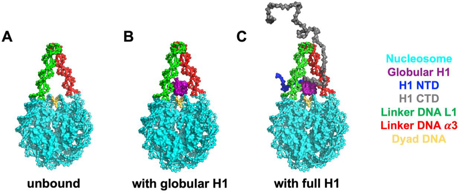
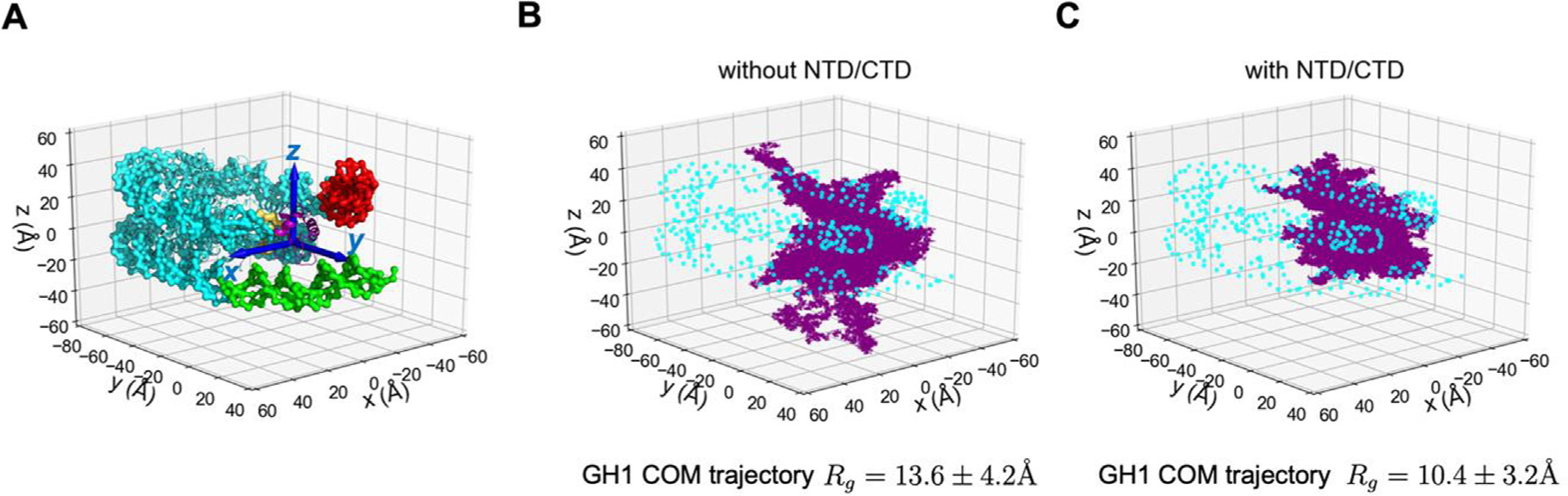
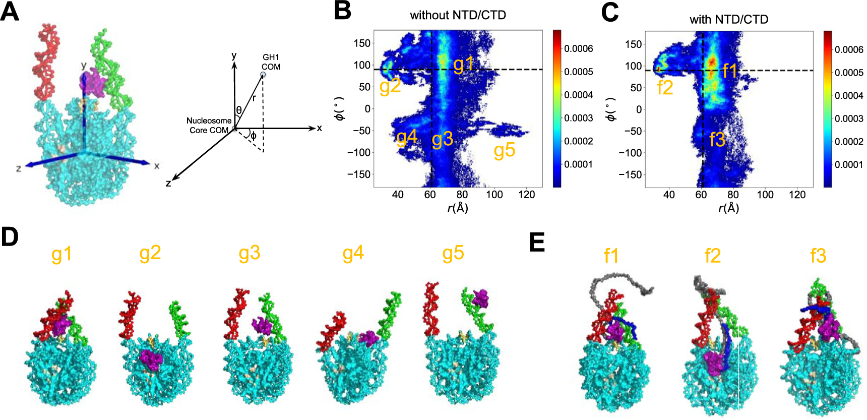
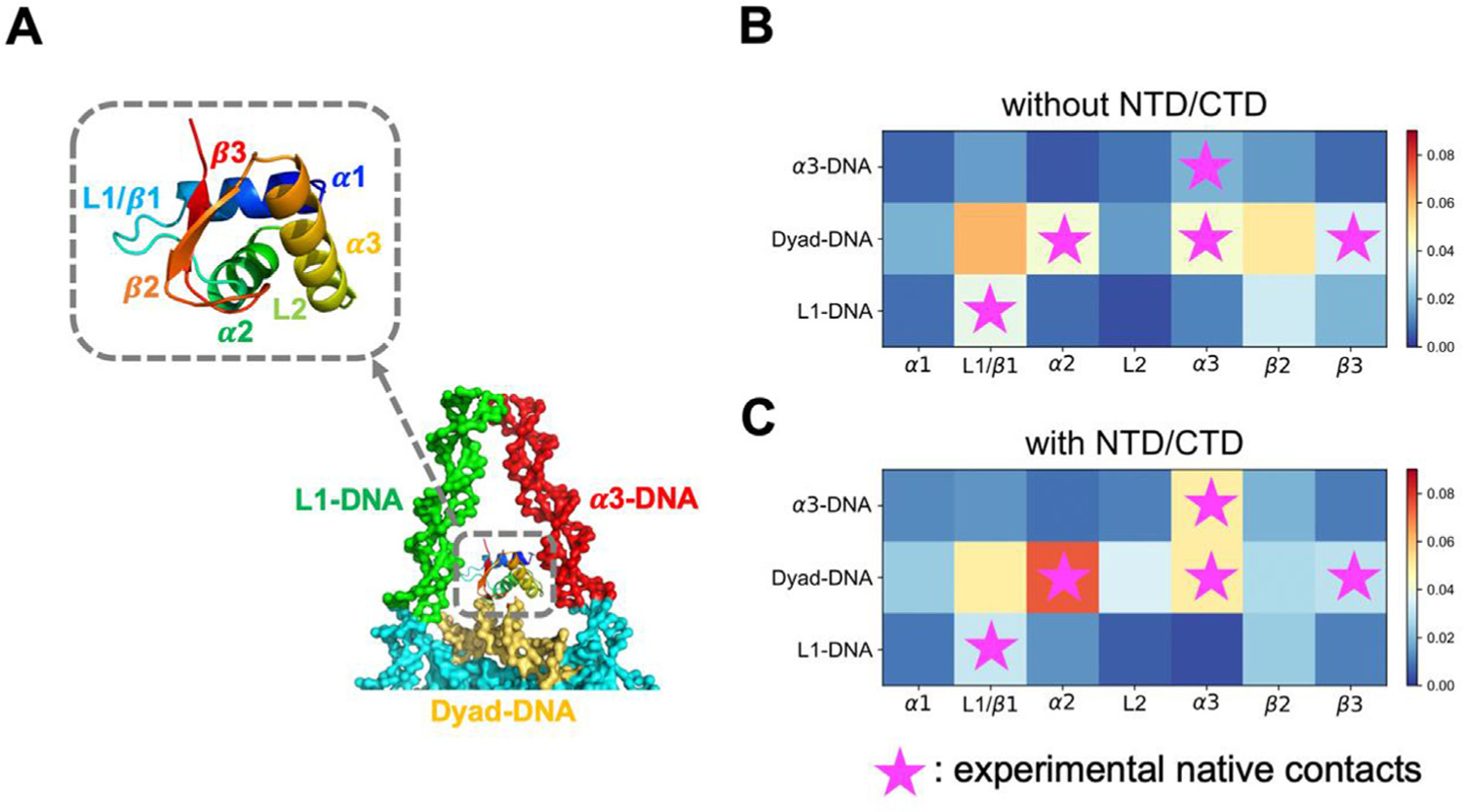
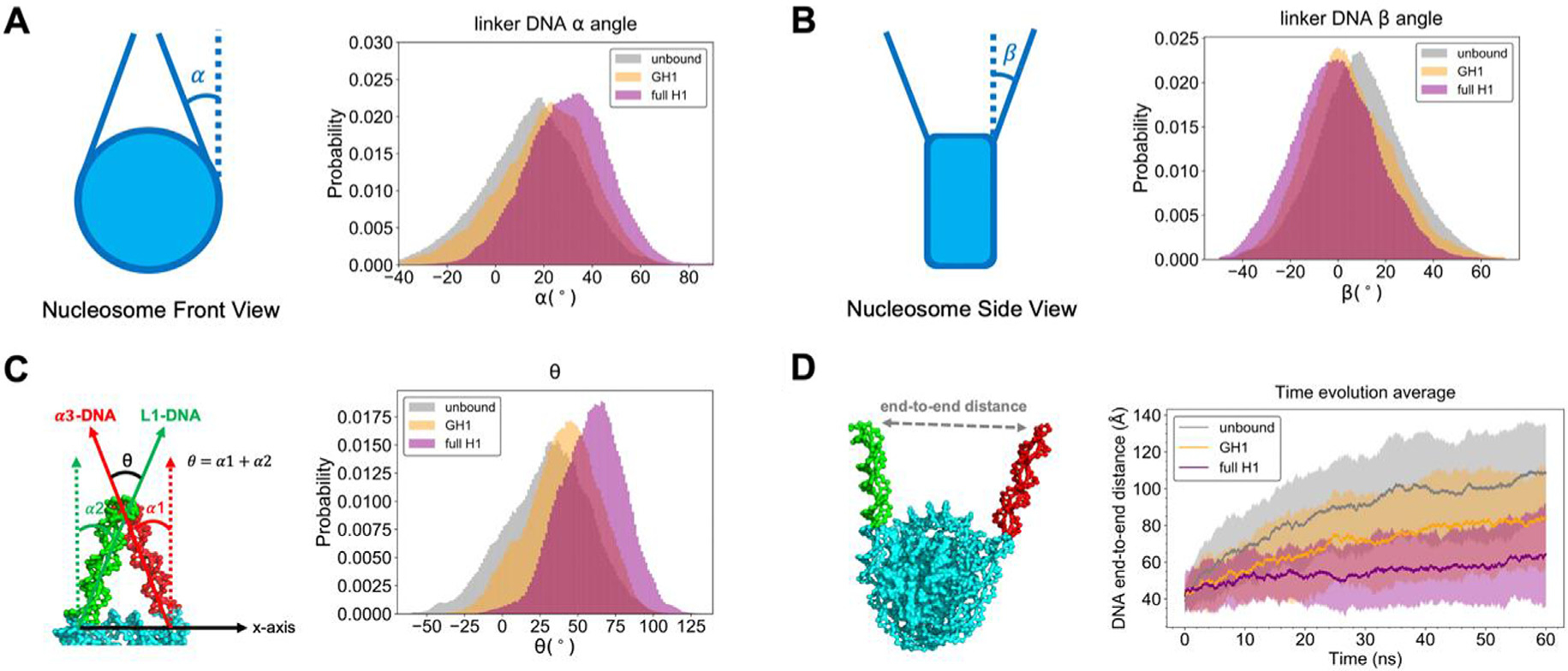
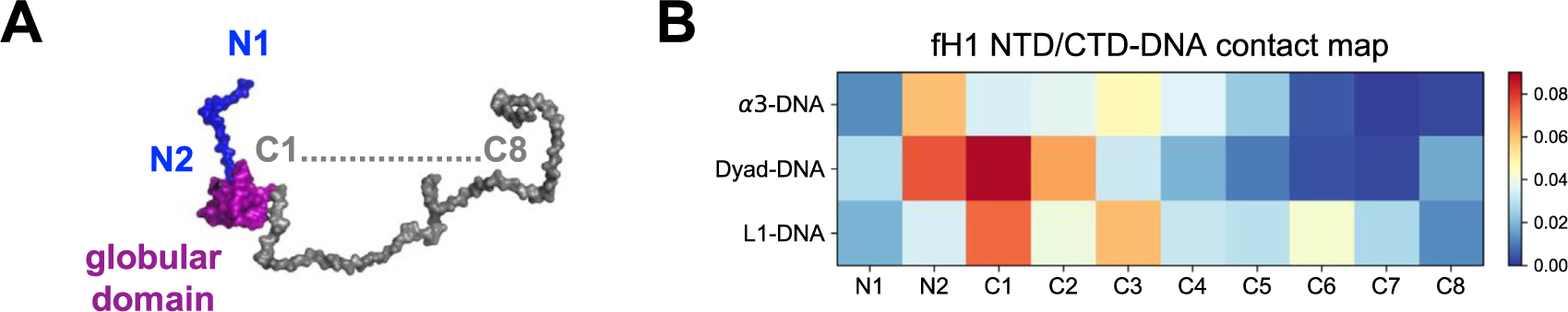
Linker histone H1 is an essential regulatory protein for many critical biological processes, such as eukaryotic chromatin packaging and gene expression. Mis-regulation of H1s is commonly observed in tumor cells, where the balance between different H1 subtypes has been shown to alter the cancer phenotype. Consisting of a rigid globular domain and two highly charged terminal domains, H1 can bind to multiple sites on a nucleosomal particle to alter chromatin hierarchical condensation levels. In particular, the disordered H1 amino- and carboxyl-terminal domains (NTD/CTD) are believed to enhance this binding affinity, but their detailed dynamics and functions remain unclear. In this work, we used a coarse-grained computational model, AWSEM-DNA, to simulate the H1.0b-nucleosome complex, namely chromatosome. Our results demonstrate that H1 disordered domains restrict the dynamics and conformation of both globular H1 and linker DNA arms, resulting in a more compact and rigid chromatosome particle. Furthermore, we identified regions of H1 disordered domains that are tightly tethered to DNA near the entry-exit site. Overall, our study elucidates at near-atomic resolution the way the disordered linker histone H1 modulates nucleosome's structural preferences and conformational dynamics.

Keywords: IDP; MD simulation; chromatin; chromatosome; protein-DNA interaction.


Conflict of interest statement

Declaration of Competing Interest The authors declare that they have no known competing financial interests or personal relationships that could have appeared to influence the work reported in this paper.

Figures

Figure 1.
Figure 1.
The molecular systems simulated in this study. From left to right: unbound nucleosome without H1 (A), globular H1-nucleosome (B) and full-length H1-nucleosome (C). All the models are based on a recent X-ray crystal structure (PDB: 5NL0) of the Xenopus laevis H1.0b-nucleosome.
Figure 2.
Figure 2.
GH1 dynamics is constrained by H1 NTD/CTD. (A) Coordinates of the chromatosome are defined by a 3D cartesian coordinate system (axes shown in blue). Different regions of the chromatosome are represented using the same color scheme as in Figure 1. The trajectories of GH1 COM in the H1-nucleosome system without H1 NTD/CTD (B) and with H1 NTD/CTD (C) are plotted as purple dots. The DNA beads are represented as cyan circles. The core histone octamer and H1 NTD/CTD are not shown in (B-C) for clarity.
Figure 3.
Figure 3.
GH1-nucleosome relative conformations. (A) Definition of the 3D reference coordinate system and GH1 COM spherical coordinates (r, θ, ϕ). The 2D histogram of (ϕ, r) for GH1-nucleosome (B) and full-length H1-nucleosome (C) are shown as heat maps. The color bar from blue to red indicates a probability from low to high. The ϕ and r values at the beginning of simulations are labeled as black dashed lines. The representative snapshots of all the major basins labeled in (B-C) are shown in (D) and (E). The color code for different parts of molecules is the same as Figure 1, except for the region in light brown for the histone acidic patch.
Figure 4.
Figure 4.
H1 disordered domains regulate and stabilize GH1-DNA binding interface. GH1 and nearby DNA structures are represented in (A), where colors represent different protein or DNA regions. (B-C) GH1-DNA contact maps without and with H1 NTD/CTD. Horizontal and vertical axes represent GH1 and DNA regions respectively. Color code from blue to red indicates the contact probability of a protein-DNA beads pair in this region from low to high. Purple stars represent the experimental “native contact regions”, where more than two residues in this GH1 region are close to a DNA region.
Figure 5.
Figure 5.
Linker DNA arms are more convergent with full-length H1 than globular H1 or unbound. The definition and histogram of α, β, and θ angles are shown in (A, B, C). (D) The time evolution of DNA end-to-end distance, where solid lines with shaded regions represent average values with standard deviations. The same set of legends is used in all the subset figures above (gray: unbound nucleosome; yellow: GH1-nucleosome; purple: full-length H1-nucleosome).
Figure 6.
Figure 6.
H1 NTD and CTD are tightly bound with DNA mainly via the residues proximal to the globular domain. (A) H1 NTD (blue) is divided into two regions (N1-N2, from GH1-distal to GH1-proximal). H1 CTD (orange) is divided into eight regions (C1-C8, from GH1-proximal to GH1-distal). (B) shows the contact map of DNA with NTD and CTD. The horizontal axis represents NTD (N1-N2) and CTD (C1-C8) regions. The vertical axis and color bar are the same as in Figure 4(B).

Similar articles

Cited by

References

    1. Kornberg RD, (1974). Chromatin structure: A repeating unit of histones and DNA. Science (80-.), 184, 868–871. 10.1126/science.184.4139.868. - DOI - PubMed
    1. Luger K, Mäder AW, Richmond RK, Sargent DF, Richmond TJ, (1997). Crystal structure of the nucleosome core particle at 2.8 Å resolution. Nature, 389, 251–260. 10.1038/38444. - DOI - PubMed
    1. Woodcock CL, Ghosh RP, (2010). Chromatin higher-order structure and dynamics. Cold Spring Harb. Perspect. Biol, 2, 10.1101/cshperspect.a000596a000596. - DOI - PMC - PubMed
    1. Takata H, Hanafusa T, Mori T, Shimura M, Iida Y, Ishikawa K, Yoshikawa K, Yoshikawa Y, Maeshima K, (2013). Chromatin compaction protects genomic DNA from radiation damage. PLoS One, 8, 1–11. 10.1371/journal.pone.0075622. - DOI - PMC - PubMed
    1. Misteli T, Gunjan A, Hock R, Bustin M, Brown DT, (2000). Dynamic binding of histone H1 to chromatin in living cells. Nature, 408, 877–881. 10.1038/35048610. - DOI - PubMed

Publication types

MeSH terms

LinkOut - more resources