. 2021 Feb 22;13(4):4926-4945.
doi: 10.18632/aging.202680. Epub 2021 Feb 22.

Functional analysis of POLD1 p.ser605del variant: the aging phenotype of MDPL syndrome is associated with an impaired DNA repair capacity

Affiliations

Functional analysis of POLD1 p.ser605del variant: the aging phenotype of MDPL syndrome is associated with an impaired DNA repair capacity

Michela Murdocca et al. Aging (Albany NY). .

Abstract

Mandibular hypoplasia, Deafness and Progeroid features with concomitant Lipodystrophy define a rare systemic disorder, named MDPL Syndrome, due to almost always a de novo variant in POLD1 gene, encoding the DNA polymerase δ. We report a MDPL female heterozygote for the recurrent p.Ser605del variant. In order to deepen the functional role of the in frame deletion affecting the polymerase catalytic site of the protein, cellular phenotype has been characterised. MDPL fibroblasts exhibit in vitro nuclear envelope anomalies, accumulation of prelamin A and presence of micronuclei. A decline of cell growth, cellular senescence and a blockage of proliferation in G0/G1 phase complete the aged cellular picture. The evaluation of the genomic instability reveals a delayed recovery from DNA induced-damage. Moreover, the rate of telomere shortening was greater in pathological cells, suggesting the telomere dysfunction as an emerging key feature in MDPL. Our results suggest an alteration in DNA replication/repair function of POLD1 as a primary pathogenetic cause of MDPL. The understanding of the mechanisms linking these cellular characteristics to the accelerated aging and to the wide spectrum of affected tissues and clinical symptoms in the MDPL patients may provide opportunities to develop therapeutic treatments for progeroid syndromes.

Keywords: DNA repair; MDPL syndrome; POLD1 gene; age-related disease; telomere damage.


Conflict of interest statement

CONFLICTS OF INTEREST: There is no conflict of interest.

The authors want to dedicate the paper to a progeroid patient who died last month.

This paper is dedicated to the memory of Walter Palone who, with his voluntary availability as a patient, has allowed us to open a path on genetic premature aging and laminopathies.

Figures

Figure 1
Figure 1
Clinical and molecular diagnosis. (A) Pictures showing patient’s clinical features at the age of 21: telangiectasia and skin scleroderma, minimal subcutaneous adipose tissue and reduced limb muscle mass. (B) MRI of the abdomen shows an over-representation of mesenteric fat. (C) Proband’ family pedigree. Full symbol indicates MDPL affected family members. (D) DNA analysis of the proband by NGS and Sanger sequencing revealed the recurrent heterozygous single amino acid in frame genetic deletion c.1812_1814delCTC, p.Ser605del in POLD1 gene, segregating as an autosomal dominant variant.
Figure 2
Figure 2
Senescence-associated β-galactosidase assay in MDPL-HDFs and WT controls. (A) Representative image of Senescence-associated β-galactosidase assay. A greater amount of intensely positive blue cells are displayed in MDPL-HDFs than in WT controls. (B) The histogram shows the average percentage of β-galactosidase-positive cells in WT (4,3%) and MDPL fibroblasts (20%). Error bars represent the SD from the analysis of 100 cells from three independent experiments and WT values are displayed as the average percentages of 2 different controls (***P < 0.001). (C) Long-term culture of WT (grey) and MDPL HDFs (black). PDL: population doubling levels.
Figure 3
Figure 3
DNA repair and protein expression after cisplatin treatment. (A) γH2AX foci in fibroblasts at different population doubling levels (PDL). (B) The graph shows the trend of γH2A.X positive cells at each time point. Error bars represent the SD from the analysis of 100 cells from three independent experiments and WT values are displayed as the average percentages of 2 different controls. (**P<0.01, ***P< 0.001). The two lines show the trend of micronuclei (light grey line) and altered nuclear morphologies (dark grey line) in MDPL-HDFs compared to WT cells, at each time point after cisplatin treatment. The percentage of micronuclei is around 2.5% in MDPL-HDFs+72h, while it decreases to ~1% in WT cells+72h (*P<0.05). After further 24h (WT and MDPL-HDFs +96h) the percentage of MN remains around 2% in MDPL-HDFs, decreasing to 0% in WT-cells (*P<0.05). Also the difference between the percentage of altered nuclear morphology is statistically significant between WT and MDPL-HDFs both at +72h and +96h (**P<0,01); (C) Western blot analysis of Polδ from MDPL and WT HDFs and following nuclear cytoplasm fractionation. Nono54 was used to check the correct fractionation and Vinculin was used as control. (F) Densitometric Analysis of Polδ nucleus/cytoplasm ratio protein levels. (**P<0.01). Western blot analysis of equal amount of total proteins from WT (D, E) and MDPL-HDFs (G, H) at h0, h24 and +24h of cisplatin treatment and following nuclear cytoplasm fractionation. (*P<0.05, **P<0.01). Vinculin was used as control. Data are presented as means ± SD.
Figure 4
Figure 4
Flow cytometry analysis of BrdU-positive cells of WT and MDPL HDFs. Representative histogram of cell cycle profiling, reporting the insets with the relative percentage of cells in different phases of cell cycle (G0/G1, S and G2/M). (I A, I B) indicate 48 hours of cell culture + 6 hours of BrdU. (III A, III B) indicate 72 hours of cell culture + 6 hours of BrdU. (II A, II B) indicate 24hours of cell culture + 24 hours of cisplatin treatment. (IV A, IV B) indicate drug effect after 24 hours from cisplatin removal.
Figure 5
Figure 5
Western blot analysis of Polδ from MDPL and WT HDFs after 1 Gy-X-irradiation. (A) Western blot and (B) densitometric analysis of Polδ nucleus/cytoplasm ratio protein levels after 1 Gy-X-irradiation in WT-HDFs. (**P<0.01). (C) Western blot and (D) densitometric analysis of Polδ nucleus/cytoplasm ratio protein levels after 1 Gy-X-irradiation in MDPL-HDFs. (**P<0.01). Data are presented as means ± SD.
Figure 6
Figure 6
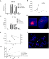
DNA repair kinetics after 1 Gy of X-irradiation. (A) DNA repair kinetics evidenced by γH2AX foci in serum-fed and serum-depleted cells after 1 Gy of X-irradiation. (B) Telomere-induced foci (TIF) in unirradiated fibroblasts at different population doubling levels (PDL). (C) Time course of TIF in serum-fed and serum-depleted cells after 1 Gy of X-irradiation. (D) Representative image of Telomeres stained with anti-TRF1 antibody (green), foci stained with anti-γH2AX antibody (red), and DAPI-stained nucleus (blue). Magnification 200X; inset shows co-localization of both antibodies, indicating a TIF. (E) Ratio between telomeric and centromeric fluorescence: T/C. (F) Representative image of chromosome spread showing telomere doublet (arrowhead) and telomere loss (arrow). Magnification 63X. (G) Telomere loss (circles, dashed lines) and telomere doublets (squares, continuous lines) at different PDL. WT in grey, MDPL in black.

References

    1. Shastry S, Simha V, Godbole K, Sbraccia P, Melancon S, Yajnik CS, Novelli G, Kroiss M, Garg A. A novel syndrome of mandibular hypoplasia, deafness, and progeroid features associated with lipodystrophy, undescended testes, and male hypogonadism. J Clin Endocrinol Metab. 2010; 95:E192–97. 10.1210/jc.2010-0419 - DOI - PMC - PubMed
    1. Cenni V, D’Apice MR, Garagnani P, Columbaro M, Novelli G, Franceschi C, Lattanzi G. Mandibuloacral dysplasia: a premature ageing disease with aspects of physiological ageing. Ageing Res Rev. 2018; 42:1–13. 10.1016/j.arr.2017.12.001 - DOI - PubMed
    1. Ajluni N, Meral R, Neidert AH, Brady GF, Buras E, McKenna B, DiPaola F, Chenevert TL, Horowitz JF, Buggs-Saxton C, Rupani AR, Thomas PE, Tayeh MK, et al.. Spectrum of disease associated with partial lipodystrophy: lessons from a trial cohort. Clin Endocrinol (Oxf). 2017; 86:698–707. 10.1111/cen.13311 - DOI - PMC - PubMed
    1. Chang LS, Zhao L, Zhu L, Chen ML, Lee MY. Structure of the gene for the catalytic subunit of human DNA polymerase delta (POLD1). Genomics. 1995; 28:411–19. 10.1006/geno.1995.1169 - DOI - PubMed
    1. Prindle MJ, Loeb LA. DNA polymerase delta in DNA replication and genome maintenance. Environ Mol Mutagen. 2012; 53:666–82. 10.1002/em.21745 - DOI - PMC - PubMed

Publication types

Supplementary concepts

LinkOut - more resources