

Comparison between conventional and compressed sensing cine cardiovascular magnetic resonance for feature tracking global circumferential strain assessment
- PMID: 33618722
- PMCID: PMC7898736
- DOI: 10.1186/s12968-021-00708-5
Comparison between conventional and compressed sensing cine cardiovascular magnetic resonance for feature tracking global circumferential strain assessment
Abstract
Background: Feature tracking (FT) has become an established tool for cardiovascular magnetic resonance (CMR)-based strain analysis. Recently, the compressed sensing (CS) technique has been applied to cine CMR, which has drastically reduced its acquisition time. However, the effects of CS imaging on FT strain analysis need to be carefully studied. This study aimed to investigate the use of CS cine CMR for FT strain analysis compared to conventional cine CMR.
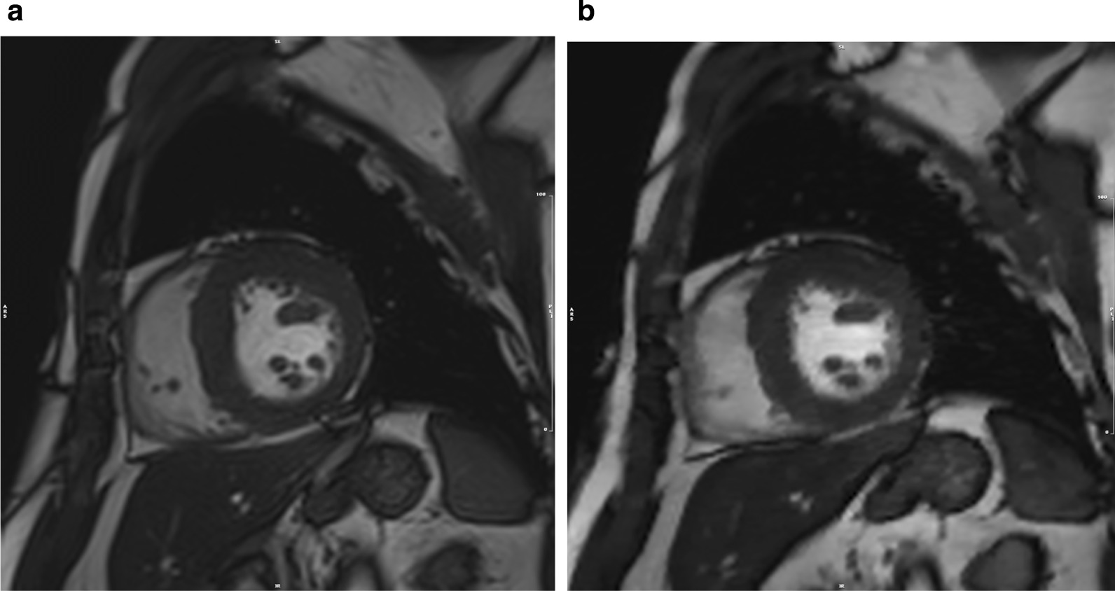
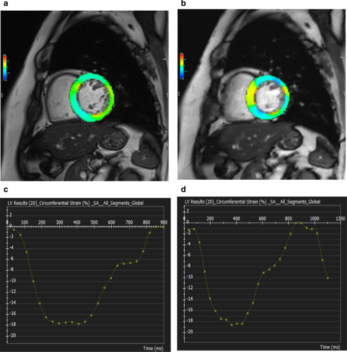
Methods: Sixty-five patients with different left ventricular (LV) pathologies underwent both retrospective conventional cine CMR and prospective CS cine CMR using a prototype sequence with the comparable temporal and spatial resolution at 3 T. Eight short-axis cine images covering the entire LV were obtained and used for LV volume assessment and FT strain analysis. Prospective CS cine CMR data over 1.5 heartbeats were acquired to capture the complete end-diastolic data between the first and second heartbeats. LV volume assessment and FT strain analysis were performed using a dedicated software (ci42; Circle Cardiovasacular Imaging, Calgary, Canada), and the global circumferential strain (GCS) and GCS rate were calculated from both cine CMR sequences.
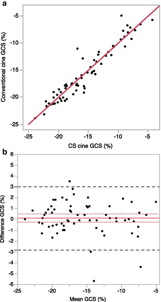
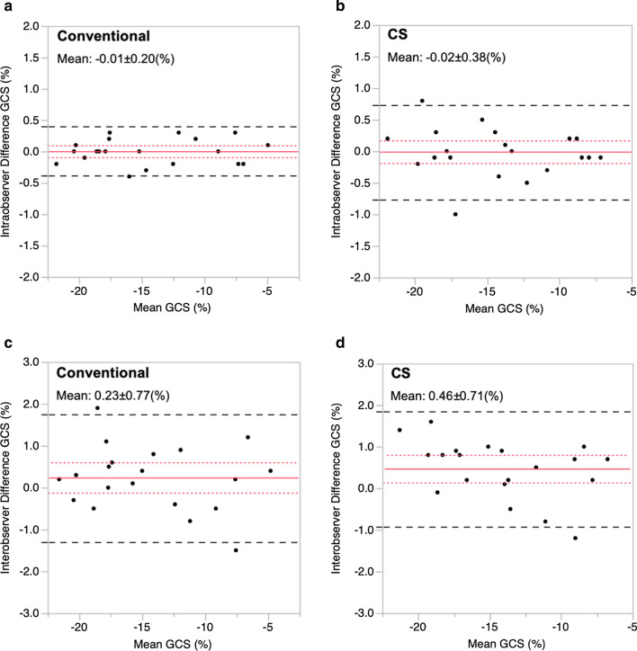
Results: There were no significant differences in the GCS (- 17.1% [- 11.7, - 19.5] vs. - 16.1% [- 11.9, - 19.3; p = 0.508) and GCS rate (- 0.8 [- 0.6, - 1.0] vs. - 0.8 [- 0.7, - 1.0]; p = 0.587) obtained using conventional and CS cine CMR. The GCS obtained using both methods showed excellent agreement (y = 0.99x - 0.24; r = 0.95; p < 0.001). The Bland-Altman analysis revealed that the mean difference in the GCS between the conventional and CS cine CMR was 0.1% with limits of agreement between -2.8% and 3.0%. No significant differences were found in all LV volume assessment between both types of cine CMR.
Conclusion: CS cine CMR could be used for GCS assessment by CMR-FT as well as conventional cine CMR. This finding further enhances the clinical utility of high-speed CS cine CMR imaging.
Keywords: Cardiovascular magnetic resonance; Compressed sensing; Feature tracking; Myocardial strain.
Conflict of interest statement
The authors declare that they have no competing interests.
Figures
Similar articles
-
Compressed sensing real-time cine cardiovascular magnetic resonance: accurate assessment of left ventricular function in a single-breath-hold.J Cardiovasc Magn Reson. 2016 Aug 24;18(1):50. doi: 10.1186/s12968-016-0271-0. J Cardiovasc Magn Reson. 2016. PMID: 27553656 Free PMC article.
-
Cardiovascular magnetic resonance feature tracking for characterization of patients with heart failure with preserved ejection fraction: correlation of global longitudinal strain with invasive diastolic functional indices.J Cardiovasc Magn Reson. 2020 Jun 4;22(1):42. doi: 10.1186/s12968-020-00636-w. J Cardiovasc Magn Reson. 2020. PMID: 32498688 Free PMC article.
-
[Feasibility of Single-Breath-Hold Compressed Sensing Real-Time Cine Imaging for Assessment of Ventricular Function and Left Ventricular Strain in Cardiac Magnetic Resonance].Sichuan Da Xue Xue Bao Yi Xue Ban. 2022 May;53(3):497-503. doi: 10.12182/20220560506. Sichuan Da Xue Xue Bao Yi Xue Ban. 2022. PMID: 35642161 Free PMC article. Chinese.
-
Principles of cardiovascular magnetic resonance feature tracking and echocardiographic speckle tracking for informed clinical use.J Cardiovasc Magn Reson. 2016 Aug 26;18(1):51. doi: 10.1186/s12968-016-0269-7. J Cardiovasc Magn Reson. 2016. PMID: 27561421 Free PMC article. Review.
-
Improving the efficiency and accuracy of cardiovascular magnetic resonance with artificial intelligence-review of evidence and proposition of a roadmap to clinical translation.J Cardiovasc Magn Reson. 2024 Winter;26(2):101051. doi: 10.1016/j.jocmr.2024.101051. Epub 2024 Jun 22. J Cardiovasc Magn Reson. 2024. PMID: 38909656 Free PMC article. Review.
Cited by
-
The effects of flip angle and gadolinium contrast agent on single breath-hold compressed sensing cardiac magnetic resonance cine for biventricular global strain assessment.Front Cardiovasc Med. 2024 Jan 29;11:1286271. doi: 10.3389/fcvm.2024.1286271. eCollection 2024. Front Cardiovasc Med. 2024. PMID: 38347952 Free PMC article.
-
Retrospective temporal resolution interpolation alters myocardial strain quantification on compressed sensing cine CMR.Int J Cardiovasc Imaging. 2025 Mar;41(3):591-602. doi: 10.1007/s10554-025-03348-3. Epub 2025 Feb 14. Int J Cardiovasc Imaging. 2025. PMID: 39953315 Free PMC article.
-
Comparison between compressed sensing and segmented cine cardiac magnetic resonance: a meta-analysis.BMC Cardiovasc Disord. 2023 Sep 21;23(1):473. doi: 10.1186/s12872-023-03426-1. BMC Cardiovasc Disord. 2023. PMID: 37735355 Free PMC article.
-
Compressed sensing cine imaging with higher temporal resolution for analysis of left atrial strain and strain rate by cardiac magnetic resonance feature tracking.Jpn J Radiol. 2023 Oct;41(10):1084-1093. doi: 10.1007/s11604-023-01433-y. Epub 2023 Apr 17. Jpn J Radiol. 2023. PMID: 37067751
-
Cardiac cine with compressed sensing real-time imaging and retrospective motion correction for free-breathing assessment of left ventricular function and strain in clinical practice.Quant Imaging Med Surg. 2023 Apr 1;13(4):2262-2277. doi: 10.21037/qims-22-596. Epub 2023 Mar 1. Quant Imaging Med Surg. 2023. PMID: 37064398 Free PMC article.
References
-
- Cicala S, de Simone G, Roman MJ, Best LG, Lee ET, Wang W, et al. Prevalence and prognostic significance of wall-motion abnormalities in adults without clinically recognized cardiovascular disease: the strong heart study. Circulation. 2007;116:143–150. doi: 10.1161/CIRCULATIONAHA.106.652149. - DOI - PubMed
-
- Vaduganathan P, He ZX, Vick GW, III, Mahmarian JJ, Verani MS. Evaluation of left ventricular wall motion, volumes, and ejection fraction by gated myocardial tomography with technetium 99 m-labeled tetrofosmin: a comparison with cine magnetic resonance imaging. J Nucl Cardiol. 1999;6:3–10. doi: 10.1016/S1071-3581(99)90058-2. - DOI - PubMed
-
- Gotte MJ, Germans T, Russel IK, Zwanenburg JJM, Marcus JT, van Rossum AC, et al. Myocardial strain and torsion quantified by cardiovascular magnetic resonance tissue tagging: studies in normal and impaired left ventricular function. J Am Coll Cardiol. 2006;48:2002–2011. doi: 10.1016/j.jacc.2006.07.048. - DOI - PubMed
Publication types
MeSH terms
LinkOut - more resources
Full Text Sources
Medical
