. 2021 Mar 2;118(9):e1916337118.
doi: 10.1073/pnas.1916337118.

Cross-reactivity of a pathogenic autoantibody to a tumor antigen in GABAA receptor encephalitis

Affiliations

Cross-reactivity of a pathogenic autoantibody to a tumor antigen in GABAA receptor encephalitis

Simone M Brändle et al. Proc Natl Acad Sci U S A. .

Abstract

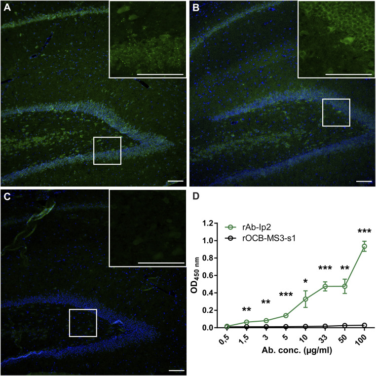
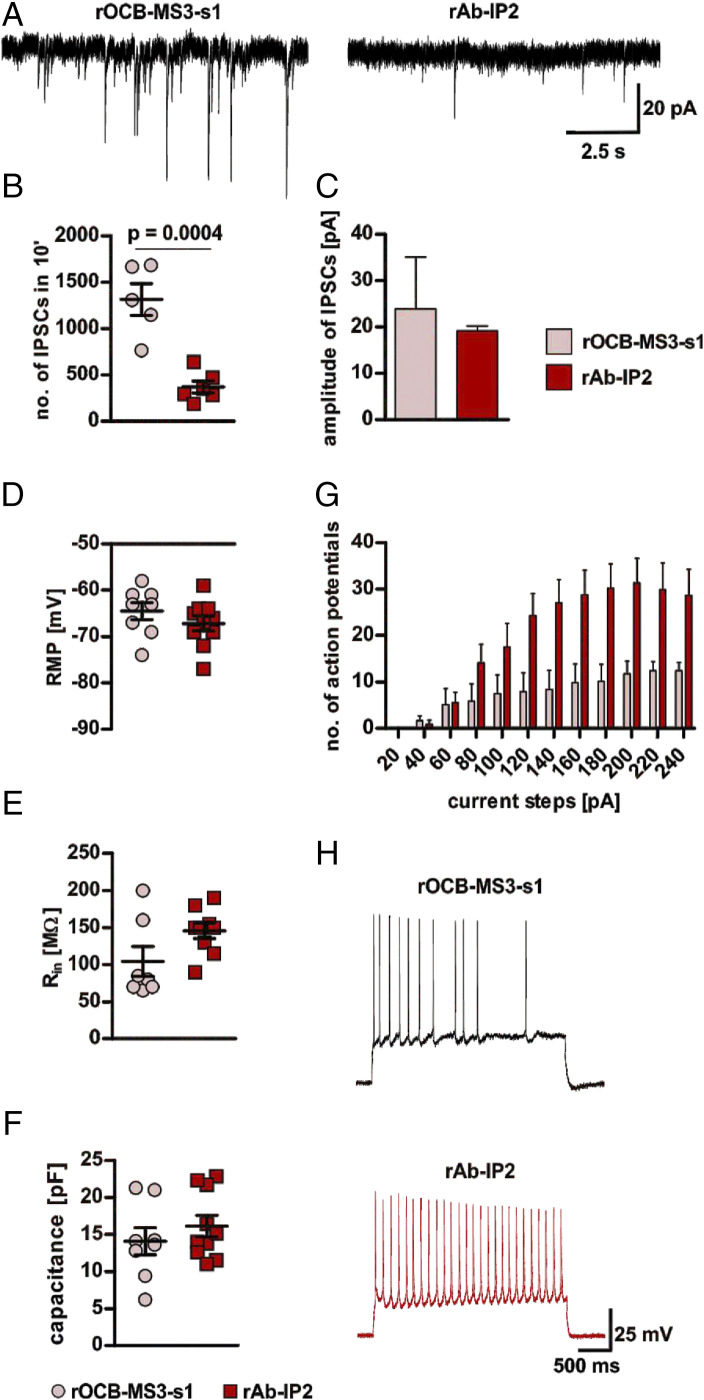
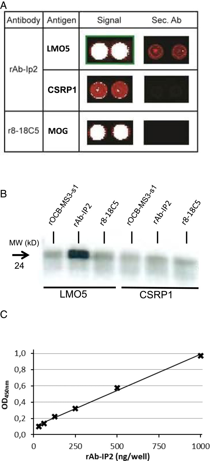
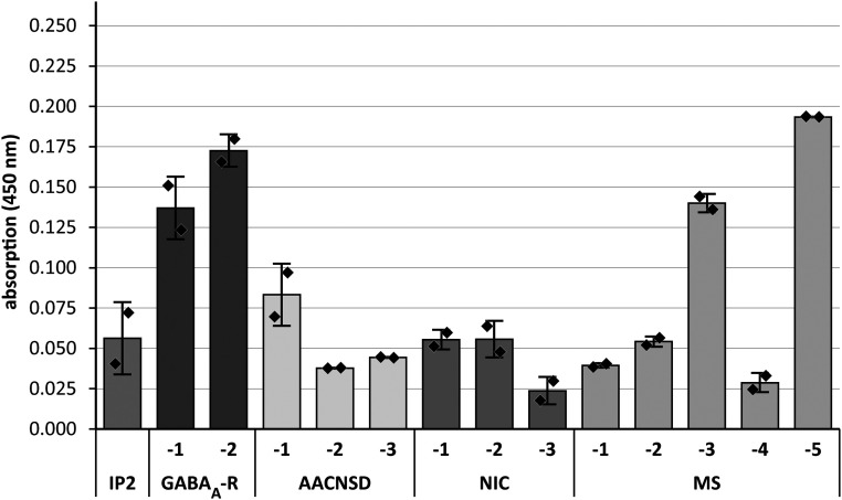
Encephalitis associated with antibodies against the neuronal gamma-aminobutyric acid A receptor (GABAA-R) is a rare form of autoimmune encephalitis. The pathogenesis is still unknown but autoimmune mechanisms were surmised. Here we identified a strongly expanded B cell clone in the cerebrospinal fluid of a patient with GABAA-R encephalitis. We expressed the antibody produced by it and showed by enzyme-linked immunosorbent assay (ELISA) and immunohistochemistry that it recognizes the GABAA-R. Patch-clamp recordings revealed that it tones down inhibitory synaptic transmission and causes increased excitability of hippocampal CA1 pyramidal neurons. Thus, the antibody likely contributed to clinical disease symptoms. Hybridization to a protein array revealed the cross-reactive protein LIM-domain-only protein 5 (LMO5), which is related to cell-cycle regulation and tumor growth. We confirmed LMO5 recognition by immunoprecipitation and ELISA and showed that cerebrospinal fluid samples from two other patients with GABAA-R encephalitis also recognized LMO5. This suggests that cross-reactivity between GABAA-R and LMO5 is frequent in GABAA-R encephalitis and supports the hypothesis of a paraneoplastic etiology.

Keywords: GABA-A-receptor encephalitis; autoantibody; autoimmune encephalitis; epilepsy; paraneoplastic encephalitis.


Conflict of interest statement

The authors declare no competing interest.

Figures

Fig. 1.
Fig. 1.
rAb-Ip2 recognizes the extracellular domain of GABAA-R-α1. Immunohistochemical staining of formalin-fixed paraffin-embedded rat hippocampus. (A) Staining with rAb-IP2 (green). Nuclei are stained with DAPI. Isotype control staining was negative. (Scale bar: 100 µm.) (B) Staining with the commercial antibody 62-3G1 to the GABAA-R-α1 subunit (green). (C) Staining with the negative control antibody rOCB-MS3-s1. (D) The ELISA shows that rAb-IP2 recognizes recombinant GABAA-R-α1ex produced in E. coli in a concentration-dependent manner (green). Control antibody rOCB-MS3-s1 does not show any reactivity to GABAA-R-α1ex (black). Error bars indicate SEM, n = 4. Statistical significance was calculated with GraphPad Prism 6 by unpaired t test. *P < 0.05,**P < 0.01,***P < 0.001.
Fig. 2.
Fig. 2.
Electrophysiological effects of rAb-IP2 on murine hippocampal CA1 pyramidal neurons. (A) Exemplary traces showing sIPSCs recorded from rOCB-MS3-s1– (control; Left) and rAb-IP2–incubated (Right) pyramidal neurons in the hippocampal region CA1. (B) Scatter plot showing that incubation with rAb-IP2 leads to a significant decrease of the number of sIPSCs recorded in an overall period of 10 min in comparison to control (rAb-IP2: 371.8 ± 65.8, n = 6; rOCB-MS3-s1: 1,316.0 ± 171.8, n = 5; unpaired Student’s t test: t = 5.511, degrees of freedom [df] = 9, P = 0.0004). (C) Bar graphs show a tendency to reduced amplitudes of sIPSCs in rAb-IP2–incubated pyramidal neurons in comparison to controls (rAb-IP2: 19.19 ± 0.60 pA, n = 3; rOCB-MS3-s1: 23.9 ± 5.6 pA, n = 4; Mann–Whitney U test: P = 0.4). (D) Scatter plot showing the RMP of rAb-IP2–incubated neurons and controls (rAbIP2: −67.20 ± 1.56, n = 10; rOCB-MS3-s1: −64.5 ± 1.83 mV, n = 8; unpaired Student’s t test: t = 1.128, df = 16, P = 0.2764). (E) Scatter plot showing the Rin of rAb-IP2–incubated neurons and controls (rAb-IP2: 145.6 ± 10.3 MΩ, n = 9; rOCB-MS3-s1: 104.3 ± 20.2 MΩ, n = 7, respectively; Mann–Whitney U test: P = 0.11). (F) Scatter plot showing the whole-cell capacitance of rAb-IP2–incubated neurons and controls (rAb-IP2: 16.12 ± 1.44 pF, n = 10; rOCB-MS3-s1: 14.07 ± 1.82 pF, n = 8, respectively; unpaired Student’s t test: t = 0.8988, df = 16, P = 0.382). (G) Bar graphs showing quantification of APs generated in response to depolarizing current steps of increasing intensity (from +20 to +240 pA, 20-pA increments, duration of 2.5 ms) in rAb-IP2–incubated pyramidal neurons and controls [mixed design two-way ANOVA, main effect of rAb-IP2- vs. rOCB-MS3-s1-incubation: F(1,165) = 6.538, P = 0.02, Sidak’s multiple comparisons test for rAb-IP2- vs. rOCB-MS3-s1: at 120 pA, P = 0.0459; at 140 pA, P = 0.0127; at 160 pA, P = 0.0115; at 180 pA, P = 0.0055; at 200 pA, P = 0.0076 and at 220 pA, P = 0.0257]. Of note, for both groups the input/output relation reflected by the proportional increase of the number of APs in response to the increasing current intensity did not differ, indicating a lower threshold for AP firing in rAb-IP2–incubated cells rather than a defective AP generation in rOCB-MS3-s1–incubated cells (main effect of input/output relation: F(11, 165) = 24,90, P < 0,0001, Sidak’s multiple comparisons test: no significance between groups). (H) Exemplary traces showing APs generated in response to a +140-pA current step at resting membrane potential in rAb-IP2–incubated (red) and rOCB-MS3-s1–incubated (control; black) pyramidal neurons.
Fig. 3.
Fig. 3.
Identification of LMO5 as antigen of Ab-IP2. (A) Hybridization of rAb-IP2 to a protein array. Sections of the developed array are shown. The first column lists the antibodies used, the second column the detected target antigens, the third column the signals of the antigens, and the fourth column the signals of the secondary antibody alone. The color code ranges from black (no reactivity) to red (medium reactivity) and white (strong reactivity). All samples were spotted in duplicate. The upper row shows the detection of LMO5 (synonym CSRP2) by rAb-IP2. The middle row shows the detection of the homologous CSRP1 by rAb-IP2. Both proteins are recognized specifically as the secondary antibody alone yields no or a much weaker signal on the array. The lowest row shows recognition of the positive control antigen major oligodendrocyte glycoprotein (MOG) by the MOG-specific antibody r8-18C5. LMO5 and MOG were detected with high affinity, whereas CSRP1 was detected with low affinity. Reprinted with permission from ref. . (B) Validation of LMO5 recognition by rAb-IP2 by immunoprecipitation. Recombinant proteins LMO5 (lanes 1–3) and CSRP1 (lanes 4–6) were produced in HEK293 cells and precipitated with antibodies rOCB-MS3-s1 (lanes 1 and 4), rAb-IP2 (lanes 2 and 5), and r8-18C5 (lanes 3 and 6). Only LMO5 could be precipitated by rAb-IP2. The blot is representative for three independent experiments. (C) Validation of LMO5 recognition by rAb-IP2 by ELISA. LMO5 was coated to plates and detected by rAb-IP2 between 0 and 1,000 µg/mL rAb-IP2 recognition of recombinant LMO5 produced in E. coli occurred in a dose-dependent manner. The shown ELISA is representative for two independent experiments. The parameter for the linear equation is y = 0,0009x + 0,0942, and the correlation coefficient is R2 = 0.9954 with P < 0.001.
Fig. 4.
Fig. 4.
Detection of anti-LMO5 antibodies in other patients and controls. CSF samples from IP2, two other patients with GABAA-R encephalitis (GABAA-R-1 and -2), three with other forms of antibody-associated CNS diseases (AACNSD-1 to -3), three with NIC (NIC-1 to -3), and five with MS (MS-1 to -5) were tested in an ELISA experiment. Clinical details are given in SI Appendix, Table S1. CSF samples were diluted 1:10. We could also detect significant signals when the samples from GABAA-R-1 and GABAA-R-2 were diluted 1:20 or 1:40, respectively. Relative absorbance units were measured as optical density at 450 nm. A representative experiment of two independent assays is shown. Assays were performed in duplicate. Error bars indicate SDs of duplicates.

References

    1. Dalmau J., Graus F., Antibody-mediated encephalitis. N. Engl. J. Med. 378, 840–851 (2018). - PubMed
    1. Melzer N., Meuth S. G., Wiendl H., Paraneoplastic and non-paraneoplastic autoimmunity to neurons in the central nervous system. J. Neurol. 260, 1215–1233 (2013). - PMC - PubMed
    1. Bost C., Pascual O., Honnorat J., Autoimmune encephalitis in psychiatric institutions: Current perspectives. Neuropsychiatr. Dis. Treat. 12, 2775–2787 (2016). - PMC - PubMed
    1. Vincent A., Bien C. G., Irani S. R., Waters P., Autoantibodies associated with diseases of the CNS: New developments and future challenges. Lancet Neurol. 10, 759–772 (2011). - PubMed
    1. van Coevorden-Hameete M. H., de Graaff E., Titulaer M. J., Hoogenraad C. C., Sillevis Smitt P. A., Molecular and cellular mechanisms underlying anti-neuronal antibody mediated disorders of the central nervous system. Autoimmun. Rev. 13, 299–312 (2014). - PubMed

Publication types

MeSH terms

LinkOut - more resources