. 2021 Mar 2;118(9):e2010213118.
doi: 10.1073/pnas.2010213118.

Unfolded and intermediate states of PrP play a key role in the mechanism of action of an antiprion chaperone

Affiliations

Unfolded and intermediate states of PrP play a key role in the mechanism of action of an antiprion chaperone

Rafayel Petrosyan et al. Proc Natl Acad Sci U S A. .

Abstract

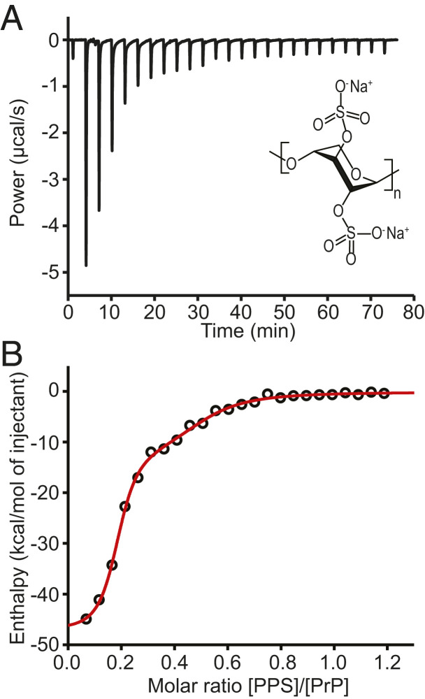
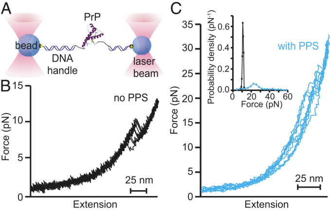
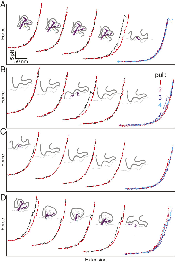
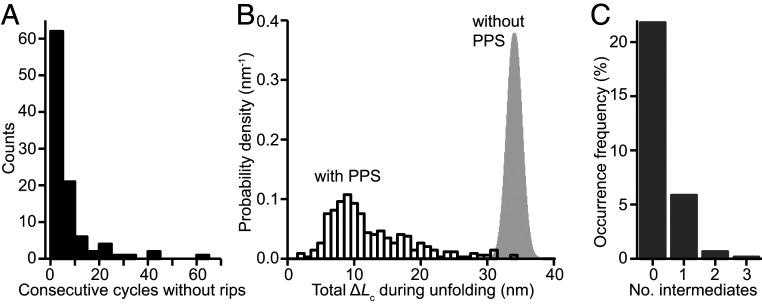
Prion and prion-like diseases involve the propagation of misfolded protein conformers. Small-molecule pharmacological chaperones can inhibit propagated misfolding, but how they interact with disease-related proteins to prevent misfolding is often unclear. We investigated how pentosan polysulfate (PPS), a polyanion with antiprion activity in vitro and in vivo, interacts with mammalian prion protein (PrP) to alter its folding. Calorimetry showed that PPS binds two sites on natively folded PrP, but one PPS molecule can bind multiple PrP molecules. Force spectroscopy measurements of single PrP molecules showed PPS stabilizes not only the native fold of PrP but also many different partially folded intermediates that are not observed in the absence of PPS. PPS also bound tightly to unfolded segments of PrP, delaying refolding. These observations imply that PPS can act through multiple possible modes, inhibiting misfolding not only by stabilizing the native fold or sequestering natively folded PrP into aggregates, as proposed previously, but also by binding to partially or fully unfolded states that play key roles in mediating misfolding. These results underline the likely importance of unfolded states as critical intermediates on the prion conversion pathway.

Keywords: energy landscape; optical tweezers; pharmacological chaperone; protein misfolding.


Conflict of interest statement

The authors declare no competing interest.

Figures

Fig. 1.
Fig. 1.
ITC of PPS binding to PrPC. (A) ITC thermogram for binding of PPS to hamster PrPC at pH 7. (Inset) Structure of PPS. (B) Binding isotherm (black) and fit to model with two independent binding sites (red).
Fig. 2.
Fig. 2.
FECs of PrP bound to PPS. (A) Schematic of force spectroscopy experiment: A single PrP molecule is attached to beads held in optical traps by DNA handles. (B) Without PPS present, PrP unfolds with a characteristic contour length change of 34.3 nm in a narrow range of forces near 10 pN. (C) With PPS present, PrP unfolds heterogeneously, showing a variety of different contour lengths and unfolding forces. (Inset) The unfolding force distribution with PPS present (cyan) is much broader than without PPS (black).
Fig. 3.
Fig. 3.
Heterogeneous unfolding of PPS-bound PrP. Four sets of successive pulling (black) and relaxing (red) curves of PrP with PPS bound show various behaviors. (Far Right) Unfolding curves for the four successive pulls. (A) PrP remains folded through the first three cycles and unfolds on the fourth. (B) PrP remains mostly unfolded but refolds between the second and third cycles. (C) PrP remains mostly unfolded after unfolding in the first cycle. (D) PrP unfolds progressively through a series of intermediates in each cycle. Insets between cycles show cartoons of PPS bound to PrP in different states.
Fig. 4.
Fig. 4.
Characteristics of FECs with PPS bound to PrP. (A) Many FECs showed no unfolding or refolding transitions. (B) The total contour-length change in FECs exhibiting transitions (black) was broadly distributed and almost always shorter than the length observed without PPS (gray). (C) A varying number of intermediates were seen for unfolding with PPS; intermediates were never seen without PPS.
Fig. 5.
Fig. 5.
Unfolding force distribution. (A) The distribution of all unfolding forces (black) does not fit well to a model that assumes a single well-defined initial state (blue), which cannot account for the long tail at high forces, but it is described well by a model allowing for heterogenous initial states (red). (B) Most of the unfolding events belong to a single cluster (cyan) defined by an unfolding force below ∼30 pN and contour length below ∼12 nm. (C) The unfolding force distribution for the dominant cluster in B is well fit by both models used in A.

Similar articles

Cited by

References

    1. Chiti F., Dobson C. M., Protein misfolding, amyloid formation, and human disease: A summary of progress over the last decade. Annu. Rev. Biochem. 86, 27–68 (2017). - PubMed
    1. Walker L. C., Jucker M., Neurodegenerative diseases: Expanding the prion concept. Annu. Rev. Neurosci. 38, 87–103 (2015). - PMC - PubMed
    1. Colby D. W., Prusiner S. B., Prions. Cold Spring Harb. Perspect. Biol. 3, a006833 (2011). - PMC - PubMed
    1. Sweeney P., et al. ., Protein misfolding in neurodegenerative diseases: Implications and strategies. Transl. Neurodegener. 6, 6 (2017). - PMC - PubMed
    1. Hartl F. U., Bracher A., Hayer-Hartl M., Molecular chaperones in protein folding and proteostasis. Nature 475, 324–332 (2011). - PubMed

Publication types

LinkOut - more resources