

Hard X-ray omnidirectional differential phase and dark-field imaging
- PMID: 33619105
- PMCID: PMC7936267
- DOI: 10.1073/pnas.2022319118
Hard X-ray omnidirectional differential phase and dark-field imaging
Abstract
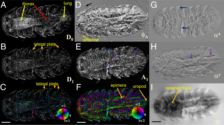
Ever since the discovery of X-rays, tremendous efforts have been made to develop new imaging techniques for unlocking the hidden secrets of our world and enriching our understanding of it. X-ray differential phase contrast imaging, which measures the gradient of a sample's phase shift, can reveal more detail in a weakly absorbing sample than conventional absorption contrast. However, normally only the gradient's component in two mutually orthogonal directions is measurable. In this article, omnidirectional differential phase images, which record the gradient of phase shifts in all directions of the imaging plane, are efficiently generated by scanning an easily obtainable, randomly structured modulator along a spiral path. The retrieved amplitude and main orientation images for differential phase yield more information than the existing imaging methods. Importantly, the omnidirectional dark-field images can be simultaneously extracted to study strongly ordered scattering structures. The proposed method can open up new possibilities for studying a wide range of complicated samples composed of both heavy, strongly scattering atoms and light, weakly scattering atoms.
Keywords: X-ray phase contrast; X-ray speckle; dark field; material science.
Copyright © 2021 the Author(s). Published by PNAS.
Conflict of interest statement
The authors declare no competing interest.
Figures


Comment in
-
On the definition, utility, and practical implementation of X-ray omnidirectional differential phase contrast and dark-field imaging.Proc Natl Acad Sci U S A. 2021 Nov 23;118(47):e2115565118. doi: 10.1073/pnas.2115565118. Proc Natl Acad Sci U S A. 2021. PMID: 34782476 Free PMC article. No abstract available.
-
Reply to Kagias and Stampanoni: High-sensitivity hard X-ray directional differential phase imaging.Proc Natl Acad Sci U S A. 2021 Nov 23;118(47):e2116067118. doi: 10.1073/pnas.2116067118. Proc Natl Acad Sci U S A. 2021. PMID: 34782478 Free PMC article. No abstract available.
Similar articles
-
2D-Omnidirectional Hard-X-Ray Scattering Sensitivity in a Single Shot.Phys Rev Lett. 2016 Mar 4;116(9):093902. doi: 10.1103/PhysRevLett.116.093902. Epub 2016 Mar 3. Phys Rev Lett. 2016. PMID: 26991177
-
Hard-X-ray directional dark-field imaging using the speckle scanning technique.Phys Rev Lett. 2015 Mar 13;114(10):103901. doi: 10.1103/PhysRevLett.114.103901. Epub 2015 Mar 11. Phys Rev Lett. 2015. PMID: 25815933
-
From synchrotron radiation to lab source: advanced speckle-based X-ray imaging using abrasive paper.Sci Rep. 2016 Feb 5;6:20476. doi: 10.1038/srep20476. Sci Rep. 2016. PMID: 26847921 Free PMC article.
-
Toward Compositional Contrast by Cryo-STEM.Acc Chem Res. 2021 Oct 5;54(19):3621-3631. doi: 10.1021/acs.accounts.1c00279. Epub 2021 Sep 7. Acc Chem Res. 2021. PMID: 34491730 Review.
-
Coherence in X-ray physics.Naturwissenschaften. 2001 Jun;88(6):249-60. doi: 10.1007/s001140100221. Naturwissenschaften. 2001. PMID: 11544952 Review.
Cited by
-
X-ray directional dark-field imaging using Unified Modulated Pattern Analysis.PLoS One. 2022 Aug 29;17(8):e0273315. doi: 10.1371/journal.pone.0273315. eCollection 2022. PLoS One. 2022. PMID: 36037163 Free PMC article.
-
Reply to Kagias and Stampanoni: High-sensitivity hard X-ray directional differential phase imaging.Proc Natl Acad Sci U S A. 2021 Nov 23;118(47):e2116067118. doi: 10.1073/pnas.2116067118. Proc Natl Acad Sci U S A. 2021. PMID: 34782478 Free PMC article. No abstract available.
-
Development of a nanometre scale X-ray speckle-based CT technique through the 3-D histological assessment of an acute respiratory distress syndrome model.Sci Rep. 2024 Oct 10;14(1):23745. doi: 10.1038/s41598-024-72660-0. Sci Rep. 2024. PMID: 39390031 Free PMC article.
-
On the definition, utility, and practical implementation of X-ray omnidirectional differential phase contrast and dark-field imaging.Proc Natl Acad Sci U S A. 2021 Nov 23;118(47):e2115565118. doi: 10.1073/pnas.2115565118. Proc Natl Acad Sci U S A. 2021. PMID: 34782476 Free PMC article. No abstract available.
-
Nano-precision metrology of X-ray mirrors with laser speckle angular measurement.Light Sci Appl. 2021 Sep 22;10(1):195. doi: 10.1038/s41377-021-00632-4. Light Sci Appl. 2021. PMID: 34552044 Free PMC article.
References
-
- Weitkamp T., et al. ., X-ray phase imaging with a grating interferometer. Opt. Express 13, 6296–6304 (2005). - PubMed
-
- Pfeiffer F., Weitkamp T., Bunk O., David C., Phase retrieval and differential phase-contrast imaging with low-brilliance X-ray sources. Nat. Phys. 2, 258–261 (2006).
-
- Pfeiffer F., et al. ., Hard-X-ray dark-field imaging using a grating interferometer. Nat. Mater. 7, 134–137 (2008). - PubMed
-
- Atsushi M., et al. ., Demonstration of X-ray Talbot interferometry. Jpn. J. Appl. Phys. 42, L866–L868 (2003).
-
- Jensen T. H., et al. ., Directional x-ray dark-field imaging of strongly ordered systems. Phys. Rev. B 82, 214103 (2010).
LinkOut - more resources
Full Text Sources
Other Literature Sources
