

Structure of 3'-PO4/5'-OH RNA ligase RtcB in complex with a 5'-OH oligonucleotide
- PMID: 33619169
- PMCID: PMC8051266
- DOI: 10.1261/rna.078692.121
Structure of 3'-PO4/5'-OH RNA ligase RtcB in complex with a 5'-OH oligonucleotide
Abstract
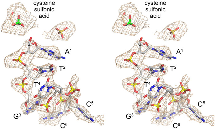
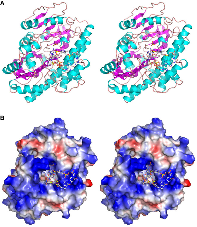
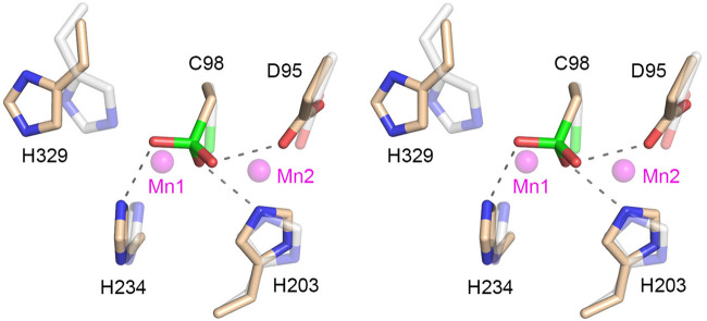
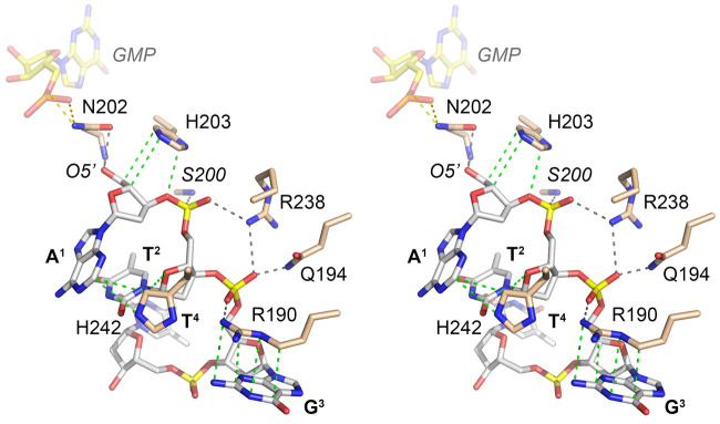
RtcB enzymes comprise a widely distributed family of manganese- and GTP-dependent RNA repair enzymes that join 2',3'-cyclic phosphate ends to 5'-OH ends via RtcB-(histidinyl-N)-GMP, RNA 3'-phosphate, and RNA3'pp5'G intermediates. RtcB can ligate either 5'-OH RNA or 5'-OH DNA strands in vitro. The nucleic acid contacts of RtcB are uncharted. Here we report a 2.7 Å crystal structure of Pyrococcus horikoshii RtcB in complex with a 6-mer 5'-OH DNA oligonucleotide HOA1pT2pG3pT4pC5pC6, which reveals enzymic contacts of Asn202 to the terminal 5'-OH nucleophile; Arg238 to the A1pT2 and T2pG3 phosphates; Arg190 and Gln194 to the T2pG3 phosphate; and an Arg190 π-cation interaction with the G3 nucleobase. The structural insights affirm functional studies of E. coli RtcB that implicated the conserved counterpart of Arg238 in engagement of the 5'-OH strand for ligation. The essential active site Cys98 that coordinates two manganese ions is oxidized to cysteine sulfonic acid in our structure, raising the prospect that RtcB activity might be sensitive to modulation during oxidative stress.
Keywords: RNA repair; cysteine sulfonic acid; tRNA splicing.
Published by Cold Spring Harbor Laboratory Press for the RNA Society.
Figures
Similar articles
-
Structures of RNA ligase RtcB in complexes with divalent cations and GTP.RNA. 2022 Nov;28(11):1509-1518. doi: 10.1261/rna.079327.122. Epub 2022 Sep 1. RNA. 2022. PMID: 36130078 Free PMC article.
-
Distinct Contributions of Enzymic Functional Groups to the 2',3'-Cyclic Phosphodiesterase, 3'-Phosphate Guanylylation, and 3'-ppG/5'-OH Ligation Steps of the Escherichia coli RtcB Nucleic Acid Splicing Pathway.J Bacteriol. 2016 Mar 31;198(8):1294-304. doi: 10.1128/JB.00913-15. Print 2016 Apr. J Bacteriol. 2016. PMID: 26858100 Free PMC article.
-
Characterization of 3'-Phosphate RNA Ligase Paralogs RtcB1, RtcB2, and RtcB3 from Myxococcus xanthus Highlights DNA and RNA 5'-Phosphate Capping Activity of RtcB3.J Bacteriol. 2015 Nov;197(22):3616-24. doi: 10.1128/JB.00631-15. Epub 2015 Sep 8. J Bacteriol. 2015. PMID: 26350128 Free PMC article.
-
Insights into the structure and function of the RNA ligase RtcB.Cell Mol Life Sci. 2023 Nov 7;80(12):352. doi: 10.1007/s00018-023-05001-5. Cell Mol Life Sci. 2023. PMID: 37935993 Free PMC article. Review.
-
RNA 3'-terminal phosphate cyclases and cyclase-like proteins.Postepy Biochem. 2016;62(3):327-334. Postepy Biochem. 2016. PMID: 28132487 Review. English.
Cited by
-
Structures of RNA ligase RtcB in complexes with divalent cations and GTP.RNA. 2022 Nov;28(11):1509-1518. doi: 10.1261/rna.079327.122. Epub 2022 Sep 1. RNA. 2022. PMID: 36130078 Free PMC article.
-
RtcB2-PrfH Operon Protects E. coli ATCC25922 Strain from Colicin E3 Toxin.Int J Mol Sci. 2022 Jun 9;23(12):6453. doi: 10.3390/ijms23126453. Int J Mol Sci. 2022. PMID: 35742896 Free PMC article.
-
Structural and biochemical characterization of the 3'-5' tRNA splicing ligases.J Biol Chem. 2025 May;301(5):108506. doi: 10.1016/j.jbc.2025.108506. Epub 2025 Apr 10. J Biol Chem. 2025. PMID: 40220997 Free PMC article.
-
Mechanistic basis for PYROXD1-mediated protection of the human tRNA ligase complex against oxidative inactivation.Nat Struct Mol Biol. 2025 Jul;32(7):1205-1212. doi: 10.1038/s41594-025-01516-6. Epub 2025 Mar 11. Nat Struct Mol Biol. 2025. PMID: 40069351 Free PMC article.
-
The life and times of a tRNA.RNA. 2023 Jul;29(7):898-957. doi: 10.1261/rna.079620.123. Epub 2023 Apr 13. RNA. 2023. PMID: 37055150 Free PMC article. Review.
References
-
- Adams PD, Afonine PV, Bunkóczi G, Chen VB, Davis IW, Echols N, Headd JJ, Hung LW, Kapral GJ, Grosse-Kunstleve RW, et al. 2010. PHENIX: a comprehensive Python-based system for macromolecular structure solution. Acta Crystrallogr D Biol Crystallogr 66: 213–221. 10.1107/S0907444909052925 - DOI - PMC - PubMed
Grants and funding
LinkOut - more resources
Full Text Sources
Other Literature Sources
Research Materials
Miscellaneous