. 2021 Feb 22;12(1):1200.
doi: 10.1038/s41467-021-21471-2.

Causal role for sleep-dependent reactivation of learning-activated sensory ensembles for fear memory consolidation

Affiliations

Causal role for sleep-dependent reactivation of learning-activated sensory ensembles for fear memory consolidation

Brittany C Clawson et al. Nat Commun. .

Abstract

Learning-activated engram neurons play a critical role in memory recall. An untested hypothesis is that these same neurons play an instructive role in offline memory consolidation. Here we show that a visually-cued fear memory is consolidated during post-conditioning sleep in mice. We then use TRAP (targeted recombination in active populations) to genetically label or optogenetically manipulate primary visual cortex (V1) neurons responsive to the visual cue. Following fear conditioning, mice respond to activation of this visual engram population in a manner similar to visual presentation of fear cues. Cue-responsive neurons are selectively reactivated in V1 during post-conditioning sleep. Mimicking visual engram reactivation optogenetically leads to increased representation of the visual cue in V1. Optogenetic inhibition of the engram population during post-conditioning sleep disrupts consolidation of fear memory. We conclude that selective sleep-associated reactivation of learning-activated sensory populations serves as a necessary instructive mechanism for memory consolidation.


Conflict of interest statement

The authors declare no competing interests.

Figures

Fig. 1
Fig. 1. Consolidation of visually cued fear memory is enhanced by post-conditioning sleep.
a At ZT0, mice underwent three stimulus–shock pairings in context A. After either 12 h of ad lib sleep or 6 h sleep deprivation (SD) followed by 6 h ad lib sleep, mice were exposed to the shock cue (X° grating) and a neutral cue (Y° grating) in context B. b Freezing behavior of the mice during the ZT12 test (Sleep: n = 19, SD: n = 16; males—solid symbols, females—open symbols). Mice allowed to sleep froze significantly more to the shock cue than mice who were sleep deprived (**p = 0.007, Holm–Sidak post hoc test). Both freely sleeping and SD mice showed higher freezing in response to the shock cue (****p < 0.0001, *p = 0.045, Holm–Sidak post hoc test; two-way RM ANOVA: main effect of sleep condition, F = 4.448, p = 0.043, main effect of orientation, F = 27.268, p < 0.0001, sleep × orientation interaction, F = 4.629, p = 0.039). c Freezing behavior quantified a discrimination index [X°/(X° + Y)] for each mouse and compared to chance performance (****p < 0.0001, Wilcoxon signed-rank test vs. chance). Values in b, c indicate mean ± SEM.
Fig. 2
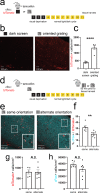
Fig. 2. TRAP labels orientation-selective V1 ensembles.
a cfos::tdTom mice were presented with either a dark screen or an oriented grating (X°) and were then injected with tamoxifen prior to 3 days of housing in complete darkness. b, c Representative V1 tdTomato labeling quantified 11 days after tamoxifen administration. ****p = 0.0001 (t = 7.07, DF = 8) for dark screen vs. X°, nested t test (n = 5 mice/condition) Values in c indicate mean ± SEM. d Prior to tissue harvest, mice were either re-exposed to gratings of the same orientation (X°) or an alternate orientation (Y°). e, f Representative images showing overlap of tdTomato (red) and cFos protein (cyan). An example of colocalization within a neuron (quantified in f) is indicated with a white arrow for each image in the inset. **p = 0.009 (t = 3.22, DF = 10), nested t test (n = 5 mice for X°, n = 7 mice for Y°). Values in f indicate mean ± SEM. g, h Densities of tdTomato+ and cFos+ neurons were similar in X°- and Y°-exposed mice. Values indicate mean ± SEM.
Fig. 3
Fig. 3. Optogenetic stimulation of TRAPed V1 neurons mimics visual experience.
a cfos::ChR2 mice with bilateral V1 fiber optics had recombination induced to a specific angle (X°). As a negative control, a second cohort was treated identically, without tamoxifen administration to induce recombination (no tamoxifen). Eleven days later, visually driven fear behavior was assessed. bd At ZT0, the mice received bilateral V1 optogenetic stimulation paired with foot shocks in lieu of the oriented grating visual stimuli used for cued conditioning in Fig. 1. At ZT12, the mice were presented with the same oriented grating used for TRAP (X°) and an alternate orientation (Y°). Optogenetically cued conditioning in tamoxifen-administered mice resulted in higher subsequent cued freezing responses to X° relative to Y° (n = 10 mice; p = 0.008 [t = 3.38, DF = 9], ratio paired t test). No tamoxifen controls showed no discrimination between X° and Y° (N.S., ratio paired t test). Values in c, d indicate mean ± SEM. eg At ZT0, a second cohort of mice underwent visually cued fear conditioning to the same orientation as the TRAPed ensemble. At ZT12, the mice received optogenetic stimulation in place of the visual cue. Freezing behavior was higher during the 3-min optogenetic stimulation than before or after stimulation in tamoxifen-administered mice (3 and 1 min, respectively; n = 5 mice; pre vs. stim—p = 0.003 [t = 7.30, DF = 4, stim vs. post—p = 0.003 [t = 7.85, DF = 4], Holm–Sidak post hoc test, one-way RM ANOVA). No tamoxifen controls showed no fear response to V1 light delivery (N.S., one-way RM ANOVA). Values in f, g indicate mean ± SEM.
Fig. 4
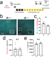
Fig. 4. TRAPed V1 neurons selective for the conditioned stimulus are reactivated in post-conditioning sleep.
a cfos::tdTom mice had recombination induced to a specific angle (X°). Eleven days later, they were cue conditioned to either the same angle as induction (X°; n = 7 mice) or an alternate angle (Y°; n = 4 mice). All mice were allowed 4.5 h of post-conditioning ad lib sleep prior to tissue harvest. b, c Representative images showing overlap of cFos expression (cyan) with tdTomato (red). The boxed region is magnified as an inset with an arrow indicating an overlapping neuron. Expression of cFos in tdTomato-labeled cells was greater for mice conditioned to the same orientation used for TRAP labeling (*p = 0.0106 [t = 2.633, DF = 63], nested t test). d, e Densities of tdTomato+ and cFos+ neurons were similar in X° and Y°-conditioned mice. Values indicate mean ± SEM.
Fig. 5
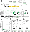
Fig. 5. Offline reactivation of orientation-selective TRAPed V1 neurons alters orientation representations in V1.
a cfos::ChR2 mice were presented with an oriented grating (X°) for TRAP. A second cohort was treated identically, without tamoxifen administration to induce recombination (no tamoxifen). Eleven days later, orientation tuning was measured repeatedly for V1 neurons recorded from anesthetized mice: at baseline, after a 20–30-min period without optogenetic stimulation, and after a 20–30-min period with 1 Hz light delivery. b Representative rasters and perievent histograms for four simultaneously recorded neurons, showing diverse firing responses during optogenetic stimulation of ChR2-expressing neurons. c The majority of stably recorded V1 neurons were reliably activated following light pulses, with variable lag times. A small proportion were inhibited by light delivery, and the remaining neurons were not affected (n = 96 neurons from 6 mice, total). Neurons recorded during rhythmic light delivery in control (no tamoxifen) mice showed no responses to light pulses (n = 79 neurons from 5 mice, total). d Power spectra for V1 LFPs showed no significant effect on ongoing rhythmic activity (N.S., K–S test, n = 5 mice). Values indicate mean ± SEM. e, f After optogenetic stimulation, neurons that were not activated following light pulses showed no change in orientation preference (N.S., nested t test, n = 37 neurons from 5 mice). In contrast, activated neurons showed increased firing rate responses for gratings of the same orientation (X°) used for TRAP. (**p = 0.004 [t = 3.93, DF = 8], nested t test, n = 40 neurons from 5 mice). g Neurons in control (no tamoxifen) mice showed no consistent orientation preference changes following rhythmic light delivery (N.S., nested t test, n = 79 neurons from 5 mice).
Fig. 6
Fig. 6. Optogenetic inhibition of orientation-selective TRAPed V1 ensembles alters orientation preference in surrounding V1 neurons.
a cfos::ArchT mice were presented with an oriented grating (X°) for TRAP. Eleven days later, V1 neurons were recorded from anesthetized mice across 30 min of optogenetic inhibition and 30 min without inhibition. Afterward, orientation preference was assessed at baseline, during a control period without optogenetic inhibition (no laser), and during a period with inhibition delivered at the same time as visual stimuli. A second cohort was treated identically, without tamoxifen administration to induce recombination (no tamoxifen). b Representative rasters and perievent histograms for 3 simultaneously recorded neurons from an Arch-expressing mouse, showing diverse firing responses during periodic optogenetic inhibition. c Distributions of stably recorded V1 neurons, which were inhibited (with >5% decrease in firing rate), activated (with >5% increase in firing rate), or unaffected by light delivery (n = 58 neurons from 5 tamoxifen treated mice, n = 278 neurons from 4 no tamoxifen control mice). d Firing rate changes with light delivery were significantly greater for Arch-expressing mice than for no tamoxifen control mice (****p = 0.0001, Mann–Whitney test). e Power spectra for V1 LFPs showed no significant change in rhythmic activity during periods of inhibition (N.S., K–S test, n = 5 mice). Values indicate mean ± SEM. f, g Neurons recorded from Arch-expressing mice that showed no decrease in firing rate during light delivery showed no change in orientation preference when light was delivered to V1 during presentation of visual stimuli (N.S., nested t test, n = 32 neurons from 5 mice). In contrast, neurons that were inhibited showed a reduced preference for gratings of the same orientation (X°) used for TRAP (**p = 0.007 [t = 3.65, DF = 8, nested t test, n = 26 neurons from 5 mice). h Neurons recorded from no tamoxifen control mice showed no consistent change in orientation preference when light was delivered to V1 during presentation of visual stimuli (N.S., nested t test, n = 52 neurons from 3 mice).
Fig. 7
Fig. 7. Sleep-specific inhibition of a V1 engram disrupts visually cued fear memory consolidation.
a cfos::ArchT mice implanted with bilateral V1 optical fibers and EEG/EMG electrodes were presented with X° for TRAP. Eleven days later, mice were conditioned using either the same orientation (X°) or an alternate orientation (Y°) as the shock cue. Post-conditioning, the mice slept with sleep-specific inhibition during the first 6 h. b No-inhibition (non-opsin-expressing) controls (n = 8) and mice cued to Y° with subsequent optogenetic inhibition (n = 8) showed higher freezing responses to the shock cue vs. the neutral cue (two-way RM ANOVA: main effect of optogenetic manipulation condition, F = 10.247, p < 0.001, main effect of orientation, F = 10.679, p = 0.004, optogenetic condition × orientation interaction, F = 7.359, p = 0.004, no-inhibition control—p = 0.002, Y°-cued inhibition—p = 0.003, Holm–Sidak post hoc test). In contrast, mice cued to X° with subsequent optogenetic inhibition (n = 8) did not differ in freezing responses to the shock cue vs. the neutral cue (N.S., Holm–Sidak post hoc test). Mice cued to either X° or Y° with subsequent inhibition showed higher freezing responses to both cues relative to no-inhibition controls, indicative of generalization. c Controls and mice cued to Y° show significant discrimination, while mice cued to X° did not (*p = 0.016 for both no-inhibition control and Y° cued with inhibition; Wilcoxon signed-rank test). Values in b, c indicate mean ± SEM.

Similar articles

Cited by

References

    1. Lutz, N. D., Diekelmann, S., Hinse-Stern, P., Born, J. & Rauss, K. Sleep supports the slow abstraction of gist from visual perceptual memories. Sci. Rep.7, 42950 (2017). - PMC - PubMed
    1. Klinzing JG, et al. Odor cueing during slow-wave sleep benefits memory independently of low cholinergic tone. Psychopharmacology. 2018;235:291–299. doi: 10.1007/s00213-017-4768-5. - DOI - PMC - PubMed
    1. Stickgold R. Parsing the role of sleep in memory processing. Curr. Opin. Neurobiol. 2013;23:847–853. doi: 10.1016/j.conb.2013.04.002. - DOI - PMC - PubMed
    1. Jones, B. J. & Spencer, R. M. C. Sleep preserves subjective and sympathetic emotional response of memories. Neurobiol. Learn. Mem. 166, 107096 (2019). - PMC - PubMed
    1. Ognjanovski N, Broussard C, Zochowski M, Aton SJ. Hippocampal network oscillations rescue memory consolidation deficits caused by sleep loss. Cereb. Cortex. 2018;28:3711–3723. doi: 10.1093/cercor/bhy174. - DOI - PMC - PubMed

Publication types