

Centriolar defects, centrin 1 alterations, and FISH studies in human spermatozoa of a male partner of a couple that produces aneuploid embryos in natural and artificial fertilization
- PMID: 33619679
- PMCID: PMC8190424
- DOI: 10.1007/s10815-021-02109-0
Centriolar defects, centrin 1 alterations, and FISH studies in human spermatozoa of a male partner of a couple that produces aneuploid embryos in natural and artificial fertilization
Abstract
Purpose: To study the potential paternal contribution to aneuploidies in the man of a couple who obtained trisomic embryos with natural and assisted fertilization.
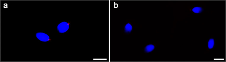
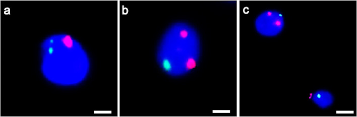
Methods: Semen analysis, immunofluorescence for localization of tubulin and centrin 1, transmission electron microscopy (TEM), and fluorescence in situ hybridization (FISH) analysis for chromosomes 18 and 9 were performed. Sperm of fertile men were used as controls.
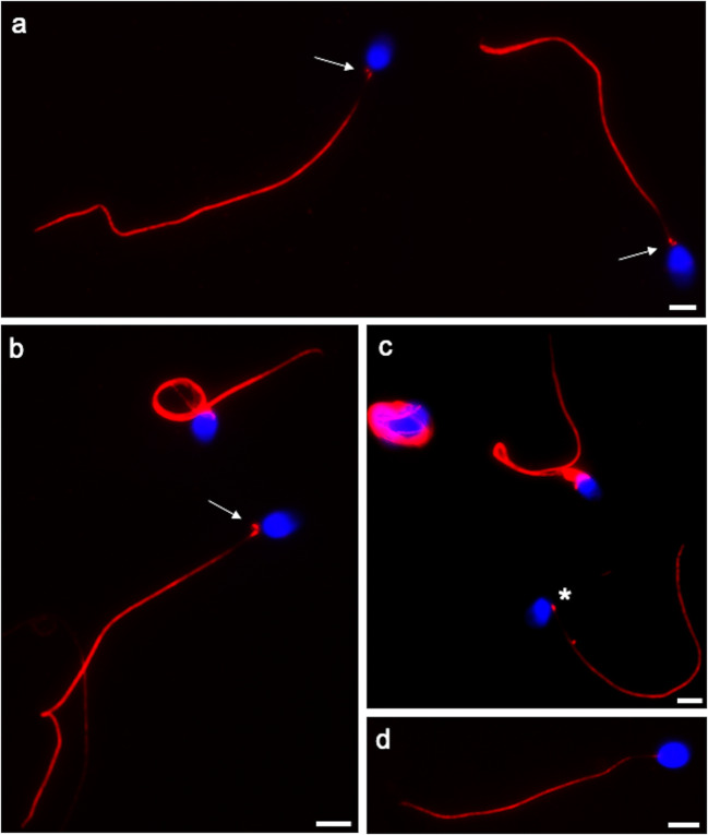
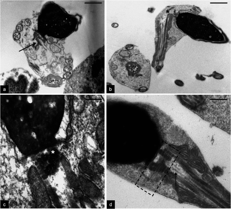
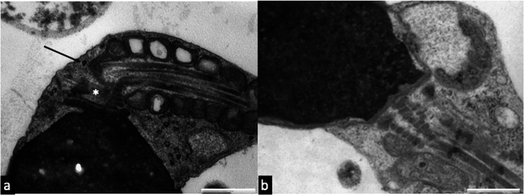
Results: The percentages of sperm motility and normal forms were decreased. The percentages of sperm with tail reduced in dimension, headless tails, coiled tails, and altered head-tail junction were significantly higher (P < 0.01) in the patient than in controls, whereas the percentage of sperm with a normal centrin 1 localization (two spots in the centriolar area) was significantly reduced (P < 0.01) in the patient. Immunofluorescence with anti-tubulin antibody showed that in most of the patient's sperm connecting pieces (83.00 ± 1.78%), two spots were present, indicating prominent proximal centriole/centriolar adjunct and evident distal centriole, whereas controls' sperm displayed a single spot, indicating the proximal centriole. The percentage of sperm with two spots was significantly higher (P < 0.01) in the patient than in controls. TEM analysis showed that centriolar adjuncts of the patient's sperm were significantly longer (721.80 ± 122.26 nm) than in controls' sperm (310.00 ± 64.11 nm; P < 0.001). The aneuploidy frequencies of the patient's sperm, detected by FISH analysis, were increased with respect to controls.
Conclusion: A paternal contribution to sperm aneuploidies cannot be excluded since the patient's sperm showed altered morphology, immature centriolar adjunct, presence of evident distal centriole, scarce presence of centrin 1, and high aneuploidy frequency.
Keywords: Aneuploidies; Centrin 1; Centriolar adjunct; FISH; Sperm; TEM.
Conflict of interest statement
The authors declare no competing interests.
Figures
Similar articles
-
Sperm with fibrous sheath dysplasia and anomalies in head-neck junction: focus on centriole and centrin 1.Andrologia. 2017 Sep;49(7). doi: 10.1111/and.12701. Epub 2016 Sep 5. Andrologia. 2017. PMID: 27596234
-
Focus on centrin in normal and altered human spermatozoa.Syst Biol Reprod Med. 2023 Jun;69(3):175-187. doi: 10.1080/19396368.2023.2181115. Epub 2023 Mar 9. Syst Biol Reprod Med. 2023. PMID: 36892570 Review.
-
Correlation between semen parameters and sperm aneuploidy rates investigated by fluorescence in-situ hybridization in infertile men.Hum Reprod. 2000 Feb;15(2):351-65. doi: 10.1093/humrep/15.2.351. Hum Reprod. 2000. PMID: 10655307
-
The sperm centriole: its inheritance, replication and perpetuation in early human embryos.Hum Reprod. 1996 Feb;11(2):345-56. doi: 10.1093/humrep/11.2.345. Hum Reprod. 1996. PMID: 8671223
-
Sperm chromosomal abnormalities and their contribution to human embryo aneuploidy.Biol Reprod. 2019 Dec 24;101(6):1091-1101. doi: 10.1093/biolre/ioz125. Biol Reprod. 2019. PMID: 31318411 Review.
Cited by
-
The relevance of sperm morphology in male infertility.Front Reprod Health. 2022 Aug 3;4:945351. doi: 10.3389/frph.2022.945351. eCollection 2022. Front Reprod Health. 2022. PMID: 36303645 Free PMC article.
-
Single cell analysis of DNA in more than 10,000 individual sperm from men with abnormal reproductive outcomes.J Assist Reprod Genet. 2021 Nov;38(11):2975-2983. doi: 10.1007/s10815-021-02300-3. Epub 2021 Aug 20. J Assist Reprod Genet. 2021. PMID: 34417660 Free PMC article.
References
MeSH terms
LinkOut - more resources
Full Text Sources
Other Literature Sources
