. 2021 Jul;239(1):125-135.
doi: 10.1111/joa.13407. Epub 2021 Feb 22.

Androgen receptors in areas of the spinal cord and brainstem: A study in adult male cats

Affiliations

Androgen receptors in areas of the spinal cord and brainstem: A study in adult male cats

Rosa L Coolen et al. J Anat. 2021 Jul.

Abstract

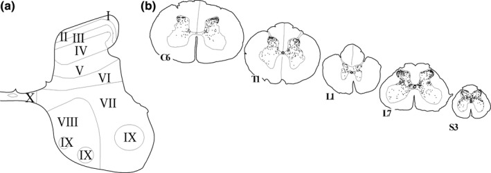
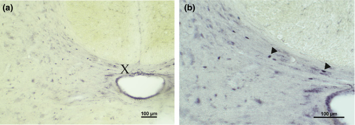
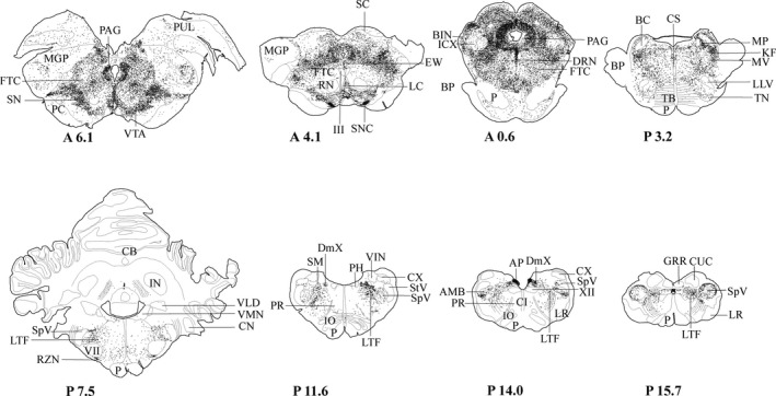
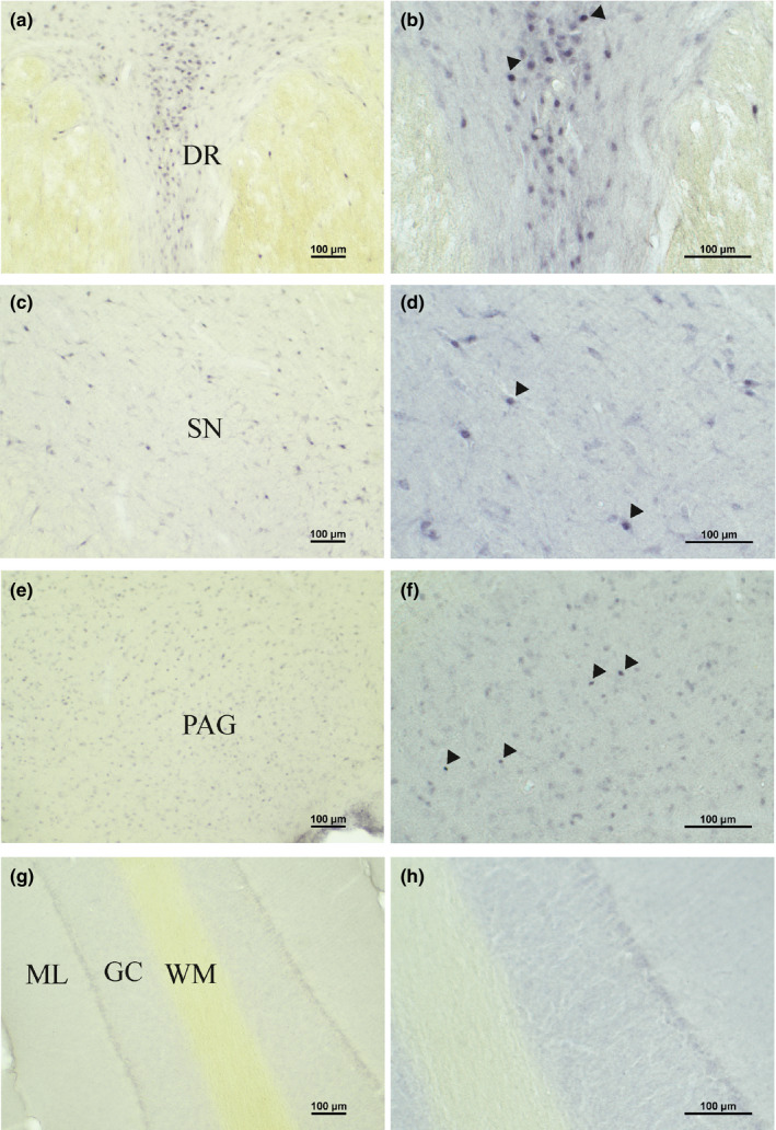
Sex hormones, including androgens and estrogens, play an important role in autonomic, reproductive and sexual behavior. The areas that are important in these behaviors lie within the spinal cord and brainstem. Relevant dysfunctional behavior in patients with altered androgen availability or androgen receptor sensitivity might be explained by the distribution of androgens and their receptors in the central nervous system. We hypothesize that autonomic dysfunction is correlated with the androgen sensitivity of spinal cord and brainstem areas responsible for autonomic functions. In this study, androgen receptor immunoreactive (AR-IR) nuclei in the spinal cord and brainstem were studied using the androgen receptor antibody PG21 in four uncastrated young adult male cats. A dense distribution of AR-IR nuclei was detected in the superior layers of the dorsal horn, including lamina I. Intensely stained nuclei, but less densely distributed, were found in lamina X and preganglionic sympathetic and parasympathetic cells of the intermediolateral cell column. Areas in the caudal brainstem showing a high density of AR-IR nuclei included the area postrema, the dorsal motor vagus nucleus and the retrotrapezoid nucleus. More cranially, the central linear nucleus in the pons contained a dense distribution of AR-IR nuclei. The mesencephalic periaqueductal gray (PAG) showed a dense distribution of AR-IR nuclei apart from the most central part of the PAG directly adjacent to the ependymal lining. Other areas in the mesencephalon with a dense distribution of AR-IR nuclei were the dorsal raphe nucleus, the retrorubral nucleus, the substantia nigra and the ventral tegmental area of Tsai. It is concluded that AR-IR nuclei are located in specific areas of the central nervous system that are involved in the control of sensory function and autonomic behavior. Furthermore, damage of these AR-IR areas might explain related dysfunction in humans.

Keywords: PG21; androgen; autonomic function; medulla oblongata; mesencephalon; spinal cord.


Conflict of interest statement

The authors declare no conflicts of interest.

Figures

FIGURE 1
FIGURE 1
AR‐IR nuclei in the spinal cord. (a) The laminae of Rexed in a schematic drawing of the 6th cervical segment. (b) Spinal cord sections (C6‐S3). Each dot represents one AR‐IR nucleus
FIGURE 2
FIGURE 2
AR‐IR nuclei in the spinal cord. Bright field photomicrographs to illustrate AR‐IR labeling in the spinal cord. Enlargement (a): ×10; (b): ×20. (a) and (b) The intermediate zone, lamina X and the central canal of spinal segment L7. X, laminae of Rexed
FIGURE 3
FIGURE 3
AR‐IR nuclei in the brainstem. Frontal brainstem sections, rostral [A6.1] to caudal [P15.7]. A6.1 corresponds with 6.1 mm anterior (A) and P15.7 corresponds with 15.7 mm posterior (P) to the frontal zero (interaural) plane. Each dot represents one AR‐IR neuron. The coordinates refer to corresponding coordinates in Berman's atlas (Berman, 1968). III, oculomotor nucleus; VII, facial nucleus; XII, hypoglossal nucleus; AMB, ambiguus nucleus; AP, area postrema; BC, brachium conjunctivum; BIN, brachial nucleus of the colliculus inferior; BP, brachium pontis; CB, cerebellum; CI, inferior central nucleus; CN, cochlear nuclei; CS, superior central nucleus; CUC, cuneate nucleus, caudal division; CX, external cuneate nucleus; DmX, dorsal motor vagus nucleus; DRN, dorsal raphe nucleus; EW, Edinger Westphal nucleus; FTC, central tegmental field; GRR, gracile nucleus, rostral division; ICX, external nucleus of the inferior colliculus; IN, nucleus interpositus; IO, inferior olive; KF, Kölliker‐Fuse nucleus; LC, central linear nucleus of the raphe; LLV, ventral nucleus of the lateral lemniscus; LR, lateral reticular nucleus; LTF, lateral tegmental field; MGP, principal nucleus of the medial geniculate body; MP, medial parabrachial nucleus; MV, motor trigeminal nucleus; P, pyramidal tract; PAG, periaqueductal gray; PC, cerebral peduncle; PH, praeposital hypoglossal nucleus; PR, paramedian reticular nucleus; PUL, pulvinar; RN, red nucleus; RZN, retrotrapezoid nucleus; SC, superior colliculus; SM, medial nucleus of the solitary tract; SN, substantia nigra; SNC, substantia nigra compact division; SpV, spinal trigeminal nucleus; StV, spinal trigeminal tract; TB, trapezoid body; TN, nucleus of the trapezoid body; VIN, inferior vestibular nucleus; VLD, lateral vestibular nucleus; VM, medial vestibular nucleus; VTA, ventral tegmental area of Tsai
FIGURE 4
FIGURE 4
AR‐IR nuclei in the brainstem. Bright field photomicrographs to illustrate AR‐IR nuclei in the brainstem. Enlargement (a, c, e, g): ×10; (b, d, f, h): ×20. (a) AR labeling in the dorsal raphe nucleus. (b) Enlargement of AR‐IR nuclei in the dorsal raphe nucleus. (c) and (d) AR‐IR nuclei in the substantia nigra. (e) and (f) AR‐IR nuclei in the PAG. (g) and (h) Absence of AR‐IR nuclei in the cerebellar cortex. DR, dorsal raphe nucleus; GC, granule cell layer; ML, molecular layer; PAG, periaqueductal gray; SN, substantia nigra; WM, white matter

Similar articles

Cited by

References

    1. Avendaño, C. & Verdu, A. (1992) Area 3a in the cat. I. A reevaluation of its location and architecture on the basis of Nissl, myelin, acetylcholinesterase, and cytochrome oxidase staining. Journal of Comparative Neurology, 321, 357–372. - PubMed
    1. Barnett, W.H. , Jenkin, S.E.M. , Milsom, W.K. , Paton, J.F.R. , Abdala, A.P. , Molkov, Y.I. , et al. (2018) The Kölliker‐Fuse nucleus orchestrates the timing of expiratory abdominal nerve bursting. Journal of Neurophysiology, 119, 401–412. - PMC - PubMed
    1. Basaria, S. , Lieb, J. , Tang, A.M. , DeWeese, T. , Carducci, M. , Eisenberger, M. , et al. (2002) Long‐term effects of androgen deprivation therapy in prostate cancer patients. Clinical Endocrinology, 56, 779–786. - PubMed
    1. Berman, A.L. (1968) The brain stem of the cat. A cytoarchitectonic atlas with stereotaxic coodinates, 1–175.
    1. Bethea, C.L. , Phu, K. , Belikova, Y. & Bethea, S.C. (2015) Localization and regulation of reproductive steroid receptors in the raphe serotonin system of male macaques. Journal of Chemical Neuroanatomy, 66, 19–27. - PMC - PubMed

Substances