Observational Study
. 2021 Aug;74(2):987-1002.
doi: 10.1002/hep.31759. Epub 2021 Jun 15.

Gene Deconvolution Reveals Aberrant Liver Regeneration and Immune Cell Infiltration in Alcohol-Associated Hepatitis

Affiliations
Observational Study

Gene Deconvolution Reveals Aberrant Liver Regeneration and Immune Cell Infiltration in Alcohol-Associated Hepatitis

Adam Kim et al. Hepatology. 2021 Aug.

Abstract

Background and aims: Acute liver damage causes hepatocyte stress and death, but in chronic liver disease impaired hepatocyte regeneration and immune cell infiltration prevents recovery. While the roles of both impaired liver regeneration and immune infiltration have been studied extensively in chronic liver diseases, the differential contribution of these factors is difficult to assess.

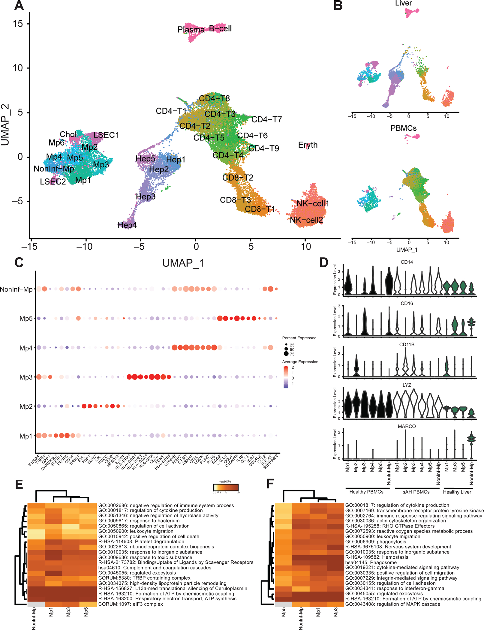
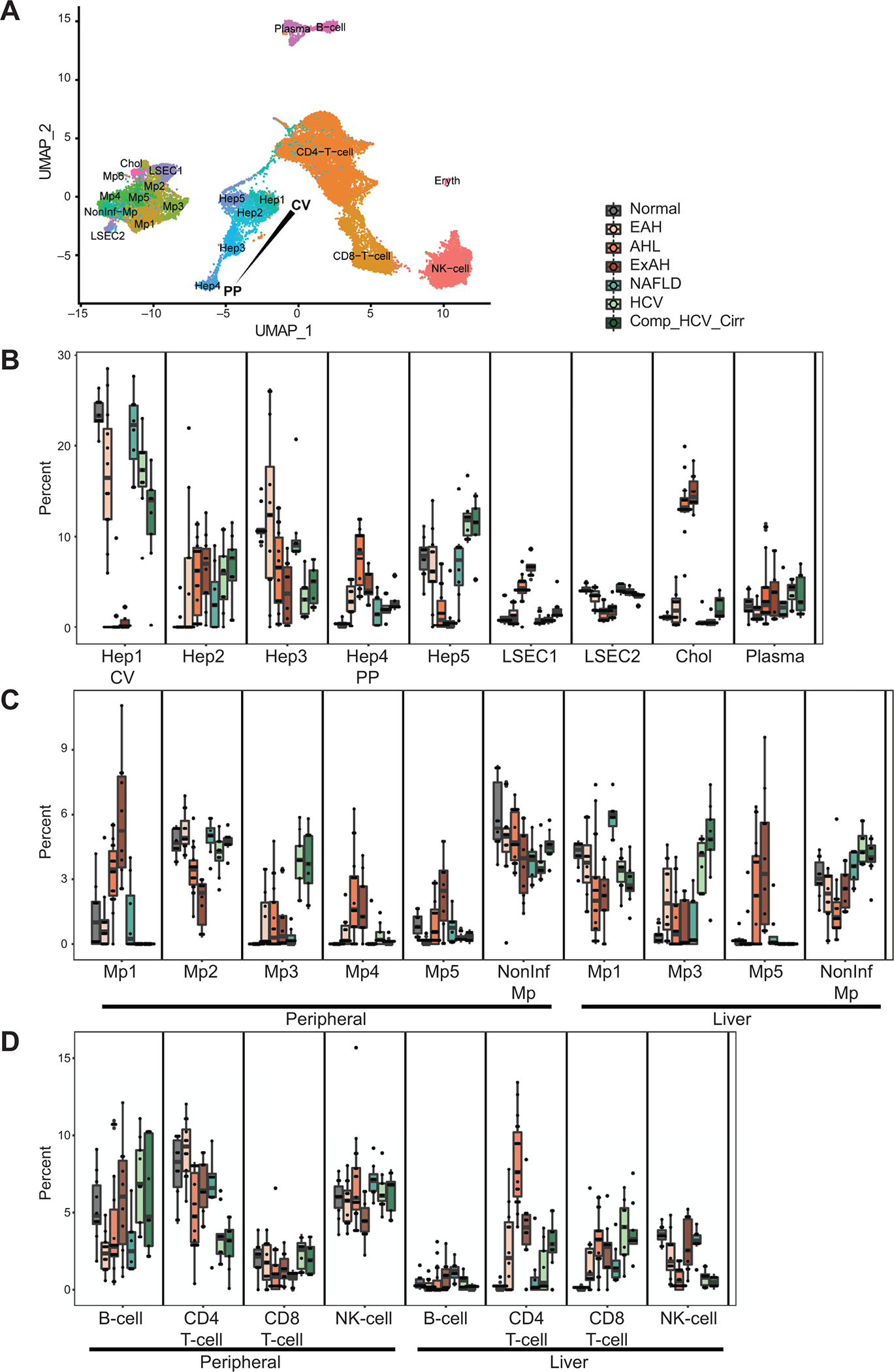
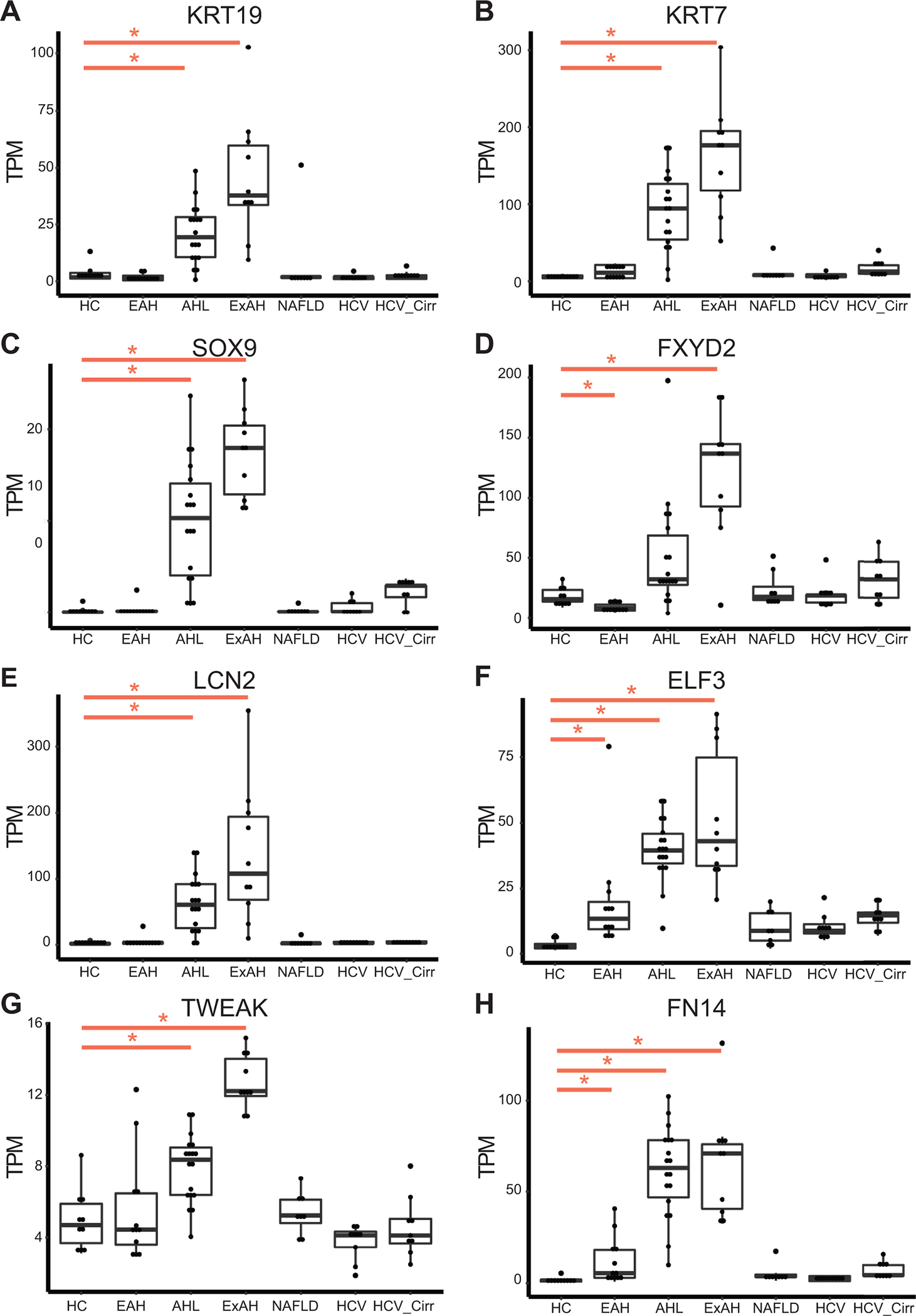
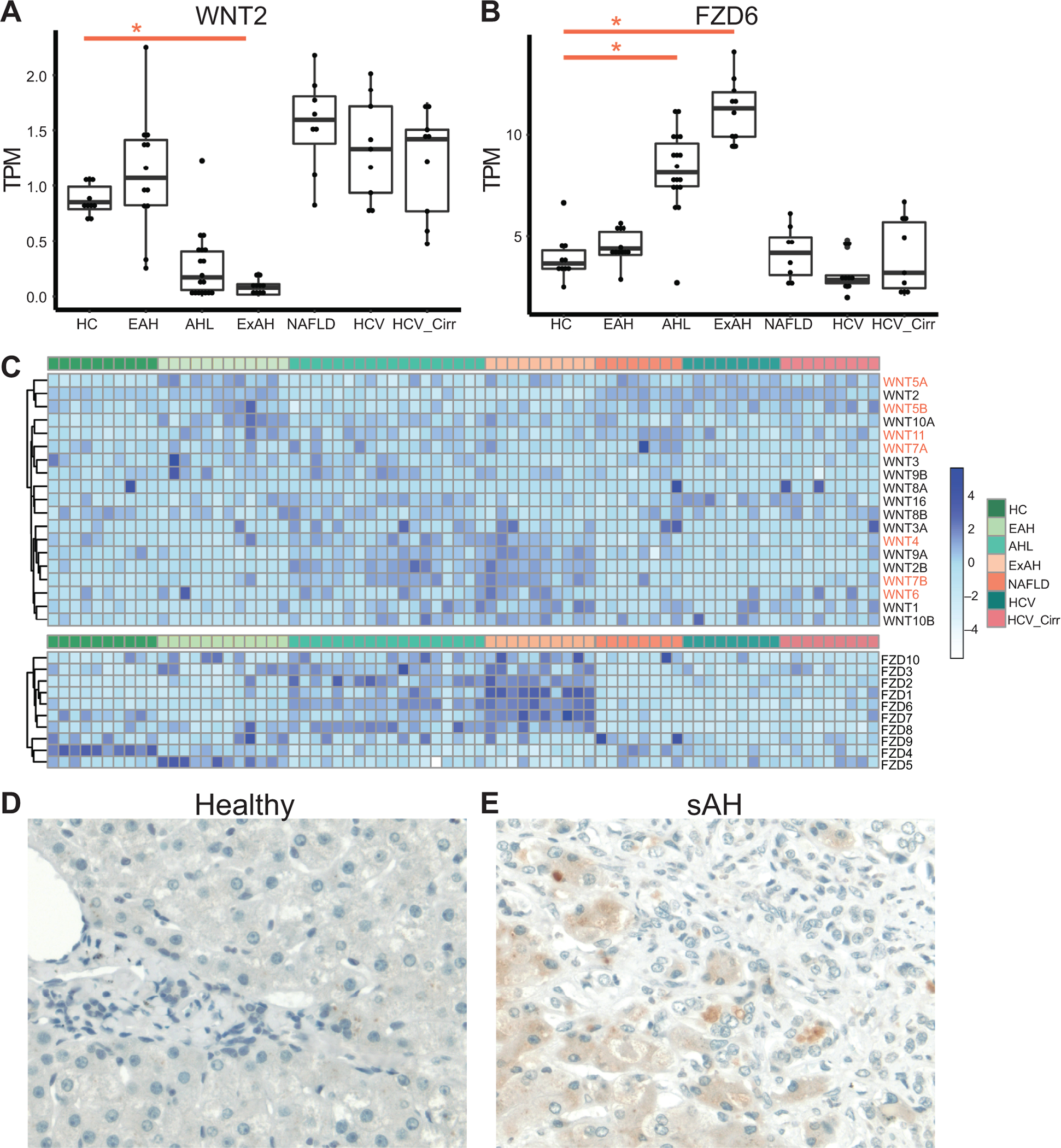
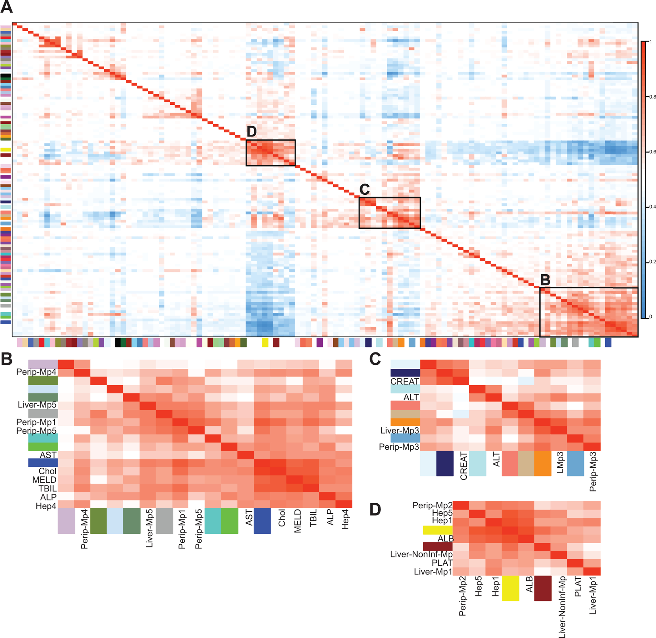
Approach and results: We combined single-cell RNA-sequencing (RNA-seq) data from healthy livers and peripheral immune cells to measure cell proportions in chronic liver diseases. Using bulk RNA-seq data from patients with early alcohol-associated hepatitis, severe AH (sAH), HCV, HCV with cirrhosis, and NAFLD, we performed gene deconvolution to predict the contribution of different cell types in each disease. Patients with sAH had the greatest change in cell composition, with increases in both periportal hepatocytes and cholangiocyte populations. Interestingly, while central vein hepatocytes were decreased, central vein endothelial cells were expanded. Endothelial cells are thought to regulate liver regeneration through WNT signaling. WNT2, important in central vein hepatocyte development, was down in sAH, while multiple other WNTs and WNT receptors were up-regulated. Immunohistochemistry revealed up-regulation of FZD6, a noncanonical WNT receptor, in hepatocytes in sAH. Immune cell populations also differed in disease. In sAH, a specific group of inflammatory macrophages was increased and distinct from the macrophage population in patients with HCV. Network and correlation analyses revealed that changes in the cell types in the liver were highly correlated with clinical liver function tests.

Conclusions: These results identify distinct changes in the liver cell populations in chronic liver disease and illustrate the power of using single-cell RNA-seq data from a limited number of samples in understanding multiple different diseases.


Conflict of interest statement

Declaration Competing of Interest

The authors declare that they have no competing of interest.

Figures

Figure 1:
Figure 1:. Combined Single-cell RNA-seq data from liver cells and PBMCs reveal similarities and differences between immune cell types.
scRNA-seq data from 5 healthy livers and PBMCs isolated from 4 sAH patients and 4 healthy controls were combined and clustered. A) UMAP of combined scRNA-seq data. B) UMAP separated by peripheral cells and liver cells. C) Dot plot showing genes upregulated in each of the 6 main myeloid-derived cell subsets, where red is higher average expression and the size of the circle represents the percentage of cells expressing. D) Violin plots showing expression of common monocyte/macrophage markers in each of the myeloid-derived clusters and separated by sample origin (healthy PBMCs, AH PBMCs, and healthy liver). E and F) Heatmap of pathway analyses showing genes up regulated in resident (E) and peripheral (F) myeloid cells.
Figure 2:
Figure 2:. Deconvolution of cell types using bulk RNA-seq data from different chronic liver diseases
Deconvolution was performed using Cibersortx and the combined scRNA-seq data which was reduced to a smaller number of clusters from both resident liver and peripheral immune cells and included in most cases 200 cells from each cluster. A) UMAP of combined scRNA-seq data after combining clusters that contained relatively few numbers of cells, including a single CD4 T-cell, CD8 T-cell, and NK-cell cluster. B-D) Proportions of each cell type in the following diseases: healthy controls (HC, n=10), early AH (EAH, n=12), AH with liver failure (AHL, n=18), explant tissue from sAH patients with emergency liver transplants (ExAH, n=10), non-alcoholic fatty liver disease (NAFLD, n=8), Hepatitis C (HCV, n=9), HCV with cirrhosis (HCV_Cirr, n=9). B) Myeloid-derived cells (MP1–5 and NonInf-Mp). C) Other immune cells including B-cells, CD4 T-cells, CD8 T-cells, and NK-cells. D) Resident liver cells, including Hep1–5, Liver Sinusoidal Endothelial Cells (LSECS1 and 2), Cholangiocytes (Chol), and Plasma cells.
Figure 3:
Figure 3:. Gene expression from different chronic liver diseases of markers of cholangiocytes and liver progenitor cells.
Boxplot of normalized gene expression levels from bulk RNA-seq data as measured by TPM (transcripts per million) for chronic liver diseases: healthy controls (HC, n=10), early AH (EAH, n=12), sAH with liver failure (AHL, n=18), explant tissue from sAH patients with emergency liver transplants (ExAH, n=10), non-alcoholic fatty liver disease (NAFLD, n=8), Hepatitis C (HCV, n=9), HCV with cirrhosis (HCV_Cirr, n=9). Significance was measured using Sleuth with (*) indicating FDR<0.05. A and B) Liver progenitor cell markers KRT7 and KRT9. C) Liver progenitor cell activation marker Sox9. D-F) Cholangiocyte specific markers FXYD2, lipocalin-2 (LCN2), E74 Like ETS Transcription Factor 3 (ELF3). G and H) TNFSF12 (TWEAK) and TNFRSF12A (TWEAK Receptor FN14).
Figure 4:
Figure 4:. Expression of WNT signaling genes in different chronic liver diseases
Boxplot of normalized gene expression levels from bulk RNA-seq data as measured by TPM (transcripts per million) for chronic liver diseases. Significance was measured using Sleuth with (*) indicating FDR<0.05. A) WNT2 expression B) FZD6 expression C) Heatmap of all WNT and FZD expression in all liver samples grouped by disease category. D and E) Immunohistochemistry using antibodies against FZD6 in paraffin-embedded sections of tissue isolated from D) healthy controls and E) patients with severe AH. Representative images are shown, n=5 per patient group.
Figure 5:
Figure 5:. Network analyses show correlation between specific cell types, gene expression, and clinical liver functional tests
WGCNA was performed to find modules of genes with highly correlated expression in the liver. These modules were then correlated to cell type proportions and clinical diagnostic measures. A) Representative heatmap of all gene modules, cell types, and different clinical measurements after hierarchical clustering. Boxed are groups of modules and clinical parameters with high correlation. B) NAFLD-associated, including modules yellow, brown, serum albumin, platelet count, peripheral Mp2, liver Mp1 and Non-Inf-Mp, Hep1 and Hep5. C) HCV-associated, including modules lightcyan1, midnightblue, paleturquoise, salmon, tan, darkorange, skyblue3, ALT, serum creatinine, and peripheral and resident Mp3. D) AH-associated, including modules thistle3, darkolivegreen4, lightsteelblue1, darkseagreen4, darkgrey, turquoise, green, blue, MELD, AST, ALP, serum bilirubin, cholangiocytes, Hep4, peripheral Mp1, 4, 5 and liver Mp5.

Comment in

References

    1. Gilgenkrantz H & Collin de l’Hortet A Understanding Liver Regeneration: From Mechanisms to Regenerative Medicine. Am J Pathol 188, 1316–1327, doi:10.1016/j.ajpath.2018.03.008 (2018). - DOI - PubMed
    1. Kubes P & Jenne C Immune Responses in the Liver. Annu Rev Immunol 36, 247–277, doi:10.1146/annurev-immunol-051116-052415 (2018). - DOI - PubMed
    1. Ben-Moshe S & Itzkovitz S Spatial heterogeneity in the mammalian liver. Nat Rev Gastroenterol Hepatol 16, 395–410, doi:10.1038/s41575-019-0134-x (2019). - DOI - PubMed
    1. Morales-Navarrete H et al.Liquid-crystal organization of liver tissue. Elife 8, doi:10.7554/eLife.44860 (2019). - DOI - PMC - PubMed
    1. Halpern KB et al.Single-cell spatial reconstruction reveals global division of labour in the mammalian liver. Nature 542, 352–356, doi:10.1038/nature21065 (2017). - DOI - PMC - PubMed

Publication types

MeSH terms