. 2021 Jul;86(1):335-345.
doi: 10.1002/mrm.28738. Epub 2021 Feb 22.

Super-resolution head and neck MRA using deep machine learning

Affiliations

Super-resolution head and neck MRA using deep machine learning

Ioannis Koktzoglou et al. Magn Reson Med. 2021 Jul.

Abstract

Purpose: To probe the feasibility of deep learning-based super-resolution (SR) reconstruction applied to nonenhanced MR angiography (MRA) of the head and neck.

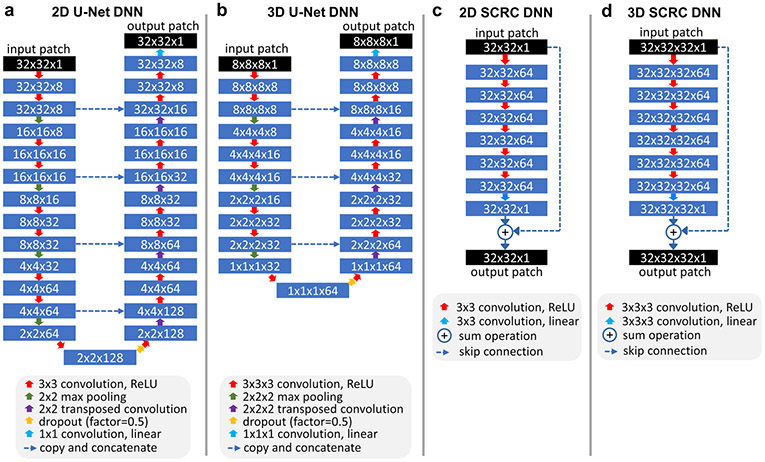
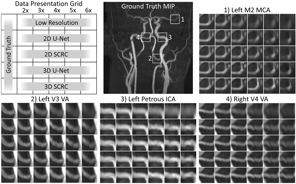
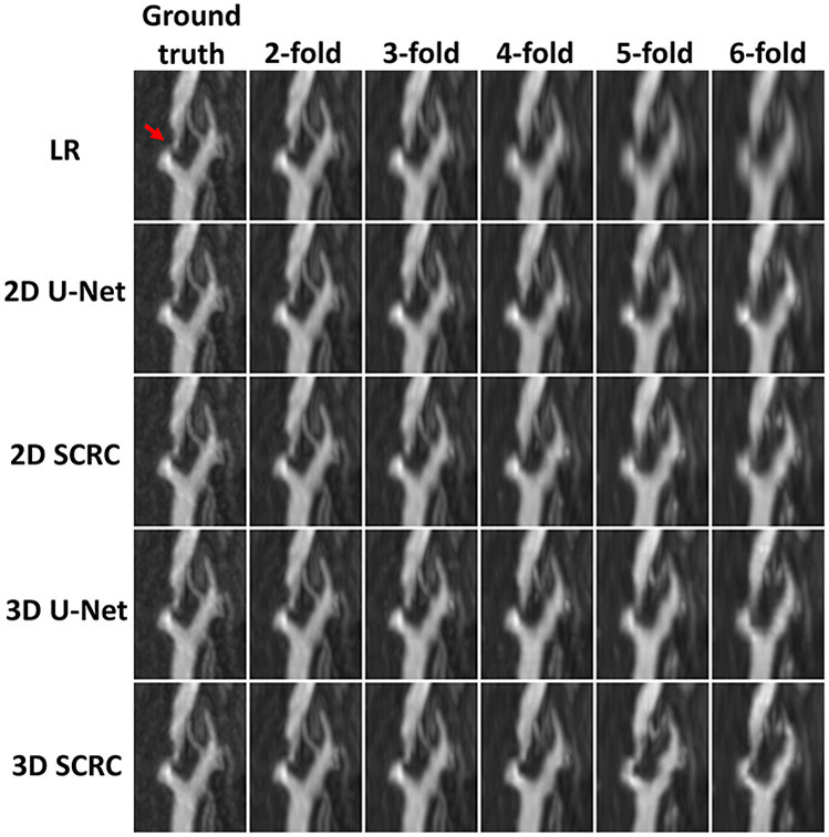
Methods: High-resolution 3D thin-slab stack-of-stars quiescent interval slice-selective (QISS) MRA of the head and neck was obtained in eight subjects (seven healthy volunteers, one patient) at 3T. The spatial resolution of high-resolution ground-truth MRA data in the slice-encoding direction was reduced by factors of 2 to 6. Four deep neural network (DNN) SR reconstructions were applied, with two based on U-Net architectures (2D and 3D) and two (2D and 3D) consisting of serial convolutions with a residual connection. SR images were compared to ground-truth high-resolution data using Dice similarity coefficient (DSC), structural similarity index measure (SSIM), arterial diameter, and arterial sharpness measurements. Image review of the optimal DNN SR reconstruction was done by two experienced neuroradiologists.

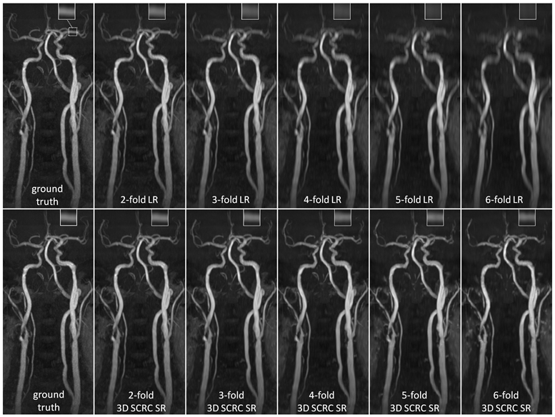
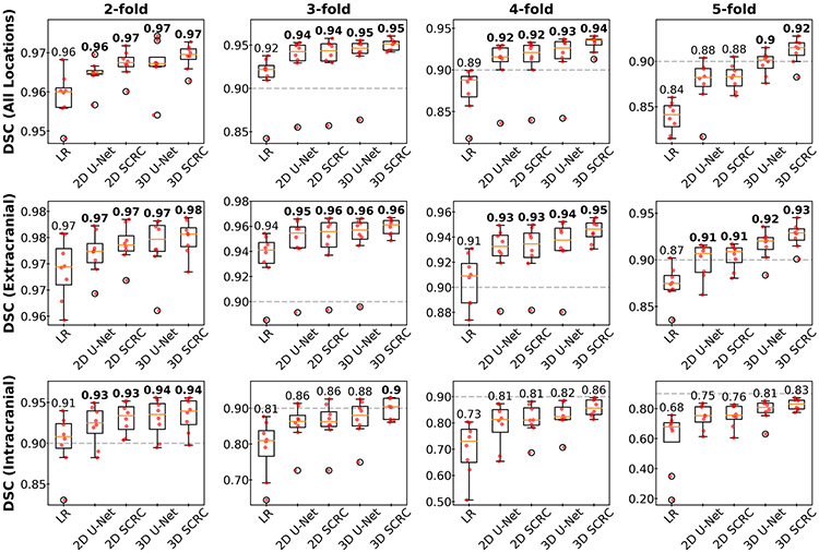
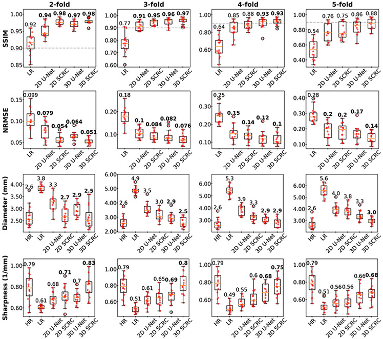
Results: DNN SR of up to twofold and fourfold lower-resolution (LR) input volumes provided images that resembled those of the original high-resolution ground-truth volumes for intracranial and extracranial arterial segments, and improved DSC, SSIM, arterial diameters, and arterial sharpness relative to LR volumes (P < .001). The 3D DNN SR outperformed 2D DNN SR reconstruction. According to two neuroradiologists, 3D DNN SR reconstruction consistently improved image quality with respect to LR input volumes (P < .001).

Conclusion: DNN-based SR reconstruction of 3D head and neck QISS MRA offers the potential for up to fourfold reduction in acquisition time for neck vessels without the need to commensurately sacrifice spatial resolution.

Keywords: MRA; deep learning; head; neck; super-resolution.


Figures

Figure 1.
Figure 1.
Architectures of the deep neural networks used for super-resolution reconstruction. ReLU = rectified linear unit.
Figure 2.
Figure 2.
Coronal maximum intensity projection 3D thin-slab stack-of-stars QISS MRA images obtained in a patient with bilateral carotid arterial disease showing the impact of 3D SCRC SR DNN reconstruction on image quality for 2- to 6-fold reduced of axial spatial resolution with respect to ground truth data (left-most column) and input lower resolution (LR) data (right-most upper panels). Insets show magnified views of the left middle cerebral artery (dashed boxed region in ground truth image). Note the improved spatial resolution of the 3D SCRC SR DNN with respect to input LR volumes as well as the improved correlation with respect to ground truth data. LR = low resolution; SCRC = serial convolution residual connection; SR = super-resolution.
Figure 3.
Figure 3.
Boxplots showing DSCs obtained with the SR DNNs for select spatial resolution reduction factors and locations. 3D DNN SR reconstructions provided the largest DSCs, with the 3D SCRC DNN providing the largest DSCs. Numbers are medians; bold numbers indicate DNNs providing DSCs≥0.9. Horizontal dashed lines show the minimum DSC adequacy threshold of 0.9. DSC = Dice similarity coefficient; LR= low resolution; SCRC = serial convolution residual connection.
Figure 4.
Figure 4.
Bar plots showing arterial SSIM, NRMSE, diameter and sharpness results obtained with the DNN SR reconstructions for select factors of spatial resolution reduction. Numbers are medians; bold numbers indicate DNNs providing SSIMs≥0.9, NRMSE values significantly differing from those of the LR volumes, and arterial sharpness and diameter measurements not significantly differing from those of the HR volumes. Dashed horizontal lines in SSIM plots indicate the minimum adequacy threshold of 0.9. SSIM = structural similarity index; NRMSE = normalized root mean square error; LR = low resolution; HR = high-resolution ground truth.
Figure 5.
Figure 5.
Comparison of DNN SR techniques in a healthy volunteer. Coronal maximum intensity projection images showing magnified views of the low resolution and DNN SR reconstructions at the four locations marked in the coronal QISS MRA image. Note the improved correlation with respect to the ground truth images obtained with the 3D DNN SR reconstructions (with respect to 2D DNN reconstructions), and with the SCRC DNNs (with respect to U-Net DNNs). Best agreement with the ground truth data was generally obtained with the 3D SCRC method. ICA = internal carotid artery; MCA = middle cerebral artery; MIP = maximum intensity projection; SCRC = serial convolution residual connection; VA = vertebral artery.
Figure 6.
Figure 6.
15-mm-thick maximum intensity projection 3D QISS MRA images obtained with the SR DNNs showing a severe stenosis of the right internal carotid artery (arrow). Note the preservation of arterial detail with the various SR DNNs for resolution reduction factors of up to ≈4. LR = low resolution; SCRC = serial convolution residual connection.

References

    1. Powers WJ, Rabinstein AA, Ackerson T, et al. Guidelines for the Early Management of Patients With Acute Ischemic Stroke: 2019 Update to the 2018 Guidelines for the Early Management of Acute Ischemic Stroke: A Guideline for Healthcare Professionals From the American Heart Association/American Stroke Association. Stroke 2019;50:e344–e418 doi: 10.1161/STR.0000000000000211. - DOI - PubMed
    1. Srinivasan A, Goyal M, Azri FA, Lum C. State-of-the-Art Imaging of Acute Stroke. RadioGraphics 2006;26:S75–S95 doi: 10.1148/rg.26si065501. - DOI - PubMed
    1. Edelman RR, Koktzoglou I. Noncontrast MR angiography: An update. J. Magn. Reson. Imaging 2019;49:355–373 doi: 10.1002/jmri.26288. - DOI - PMC - PubMed
    1. Koktzoglou I, Huang R, Ong AL, Aouad PJ, Walker MT, Edelman RR. High spatial resolution whole-neck MR angiography using thin-slab stack-of-stars quiescent interval slice-selective acquisition. Magn. Reson. Med 2020;84:3316–3324 doi: 10.1002/mrm.28339. - DOI - PMC - PubMed
    1. Saver Jeffrey L Time Is Brain—Quantified. Stroke 2006;37:263–266 doi: 10.1161/01.STR.0000196957.55928.ab. - DOI - PubMed

Publication types