

Crystal structure of acetoacetyl-CoA reductase from Rickettsia felis
- PMID: 33620038
- PMCID: PMC7900926
- DOI: 10.1107/S2053230X21001497
Crystal structure of acetoacetyl-CoA reductase from Rickettsia felis
Abstract
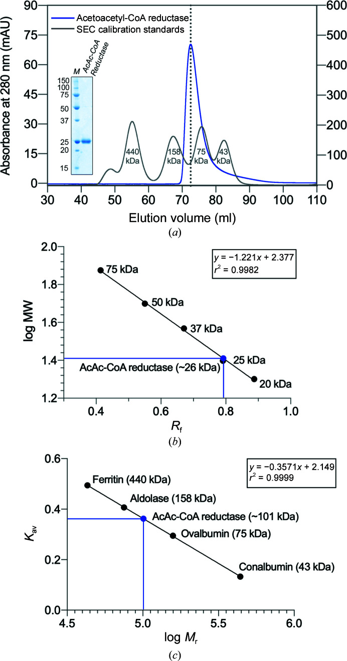
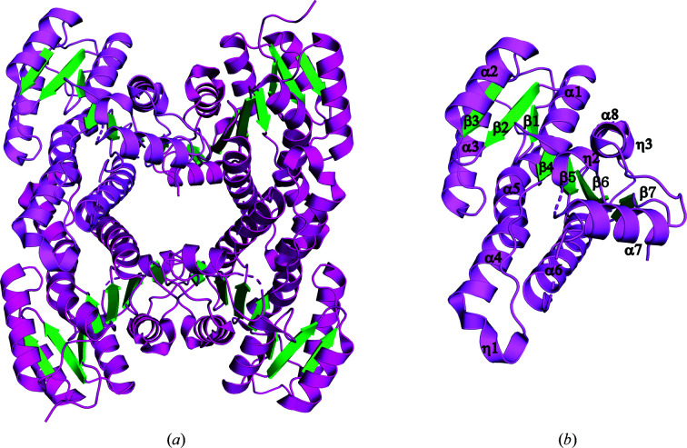
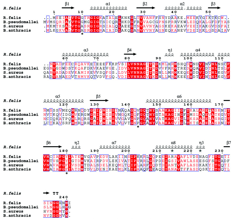
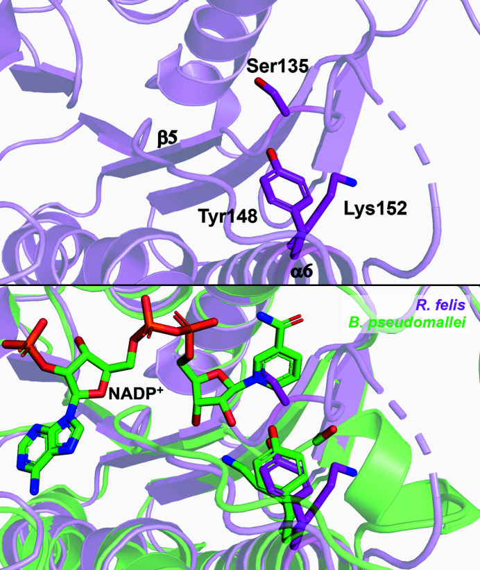
Rickettsia felis, a Gram-negative bacterium that causes spotted fever, is of increasing interest as an emerging human pathogen. R. felis and several other Rickettsia strains are classed as National Institute of Allergy and Infectious Diseases priority pathogens. In recent years, R. felis has been shown to be adaptable to a wide range of hosts, and many fevers of unknown origin are now being attributed to this infectious agent. Here, the structure of acetoacetyl-CoA reductase from R. felis is reported at a resolution of 2.0 Å. While R. felis acetoacetyl-CoA reductase shares less than 50% sequence identity with its closest homologs, it adopts a fold common to other short-chain dehydrogenase/reductase (SDR) family members, such as the fatty-acid synthesis II enzyme FabG from the prominent pathogens Staphylococcus aureus and Bacillus anthracis. Continued characterization of the Rickettsia proteome may prove to be an effective means of finding new avenues of treatment through comparative structural studies.
Keywords: PhaB; Rickettsia; SSGCID; Seattle Structural Genomics Center for Infectious Disease; acetoacetyl-CoA reductase; oxidoreductase; structural genomics.
Figures
Similar articles
-
Structure-directed construction of a high-performance version of the enzyme FabG from the photosynthetic microorganism Synechocystis sp. PCC 6803.FEBS Lett. 2015 Oct 7;589(20 Pt B):3052-7. doi: 10.1016/j.febslet.2015.09.001. Epub 2015 Sep 7. FEBS Lett. 2015. PMID: 26358291
-
Binding of NADP+ triggers an open-to-closed transition in a mycobacterial FabG β-ketoacyl-ACP reductase.Biochem J. 2017 Mar 7;474(6):907-921. doi: 10.1042/BCJ20161052. Biochem J. 2017. PMID: 28126742
-
Directed evolution and structural analysis of NADPH-dependent Acetoacetyl Coenzyme A (Acetoacetyl-CoA) reductase from Ralstonia eutropha reveals two mutations responsible for enhanced kinetics.Appl Environ Microbiol. 2013 Oct;79(19):6134-9. doi: 10.1128/AEM.01768-13. Epub 2013 Aug 2. Appl Environ Microbiol. 2013. PMID: 23913421 Free PMC article.
-
Rickettsia felis: from a rare disease in the USA to a common cause of fever in sub-Saharan Africa.Clin Microbiol Infect. 2011 Jul;17(7):996-1000. doi: 10.1111/j.1469-0691.2011.03516.x. Clin Microbiol Infect. 2011. PMID: 21722253 Review.
-
Rickettsia felis: The Complex Journey of an Emergent Human Pathogen.Trends Parasitol. 2016 Jul;32(7):554-564. doi: 10.1016/j.pt.2016.04.009. Epub 2016 May 4. Trends Parasitol. 2016. PMID: 27155905 Review.
Cited by
-
Structural and functional characterization of FabG4 from Mycolicibacterium smegmatis.Acta Crystallogr F Struct Biol Commun. 2024 Apr 1;80(Pt 4):82-91. doi: 10.1107/S2053230X2400356X. Epub 2024 Apr 24. Acta Crystallogr F Struct Biol Commun. 2024. PMID: 38656226 Free PMC article.
-
Characterization of a family I inorganic pyrophosphatase from Legionella pneumophila Philadelphia 1.Acta Crystallogr F Struct Biol Commun. 2023 Oct 1;79(Pt 10):257-266. doi: 10.1107/S2053230X23008002. Epub 2023 Sep 20. Acta Crystallogr F Struct Biol Commun. 2023. PMID: 37728609 Free PMC article.
-
Carbohydrate Deacetylase Unique to Gut Microbe Bacteroides Reveals Atypical Structure.Biochemistry. 2025 Jan 7;64(1):180-191. doi: 10.1021/acs.biochem.4c00519. Epub 2024 Dec 11. Biochemistry. 2025. PMID: 39663570 Free PMC article.
-
Structural characterization of dUTPase from Legionella pneumophila.Acta Crystallogr F Struct Biol Commun. 2025 Apr 1;81(Pt 4):155-162. doi: 10.1107/S2053230X25001815. Epub 2025 Mar 17. Acta Crystallogr F Struct Biol Commun. 2025. PMID: 40091853
-
Structures of Legionella pneumophila serogroup 1 peptide deformylase bound to nickel(II) and actinonin.Acta Crystallogr F Struct Biol Commun. 2025 Apr 1;81(Pt 4):163-170. doi: 10.1107/S2053230X25001876. Epub 2025 Mar 17. Acta Crystallogr F Struct Biol Commun. 2025. PMID: 40091854
References
-
- Alexandrov, A., Vignali, M., LaCount, D. J., Quartley, E., de Vries, C., De Rosa, D., Babulski, J., Mitchell, S. F., Schoenfeld, L. W., Fields, S., Hol, W. G. J., Dumont, M. E., Phizicky, E. M. & Grayhack, E. J. (2004). Mol. Cell. Proteomics, 3, 934–938. - PubMed
-
- Angelakis, E., Mediannikov, O., Parola, P. & Raoult, D. (2016). Trends Parasitol. 32, 554–564. - PubMed
MeSH terms
Substances
Grants and funding
LinkOut - more resources
Full Text Sources
Other Literature Sources
Miscellaneous
