. 2021 Feb;1(2):e38.
doi: 10.1002/cpz1.38.

Single-Cell RNA Sequencing Analysis of the Drosophila Larval Ventral Cord

Affiliations

Single-Cell RNA Sequencing Analysis of the Drosophila Larval Ventral Cord

Tho Huu Nguyen et al. Curr Protoc. 2021 Feb.

Erratum in

Abstract

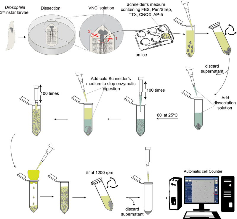
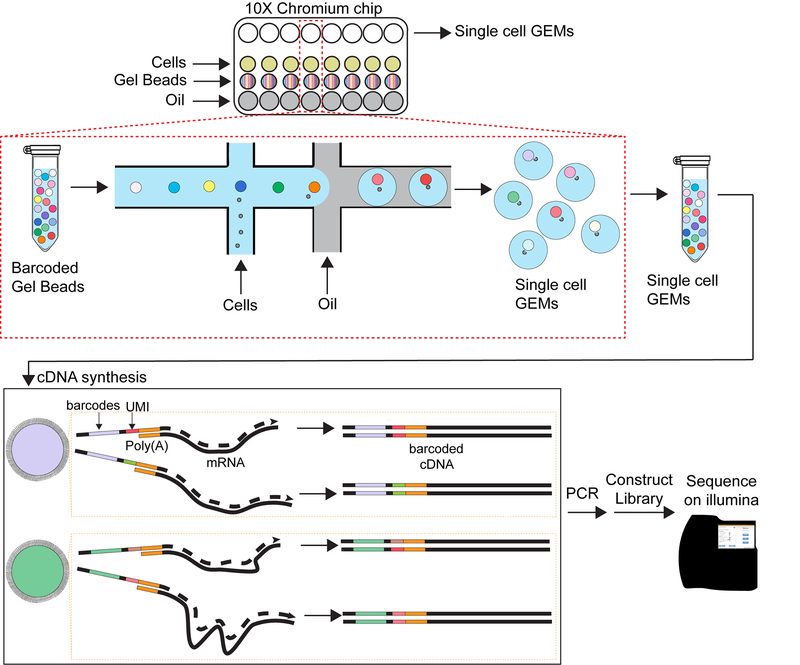
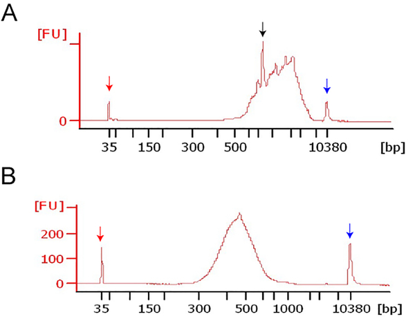
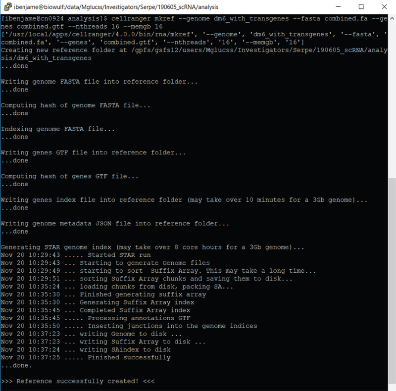
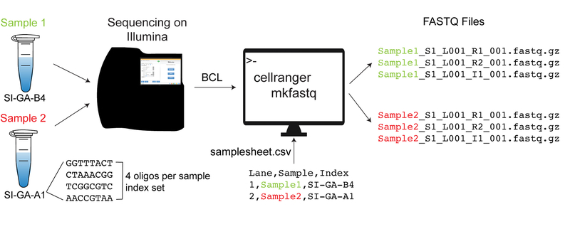
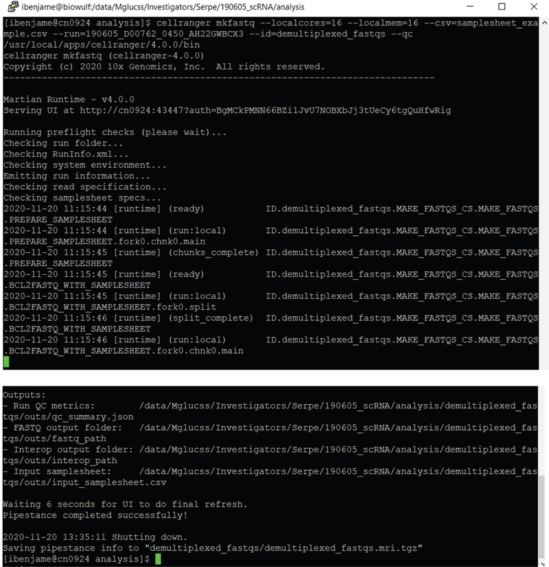
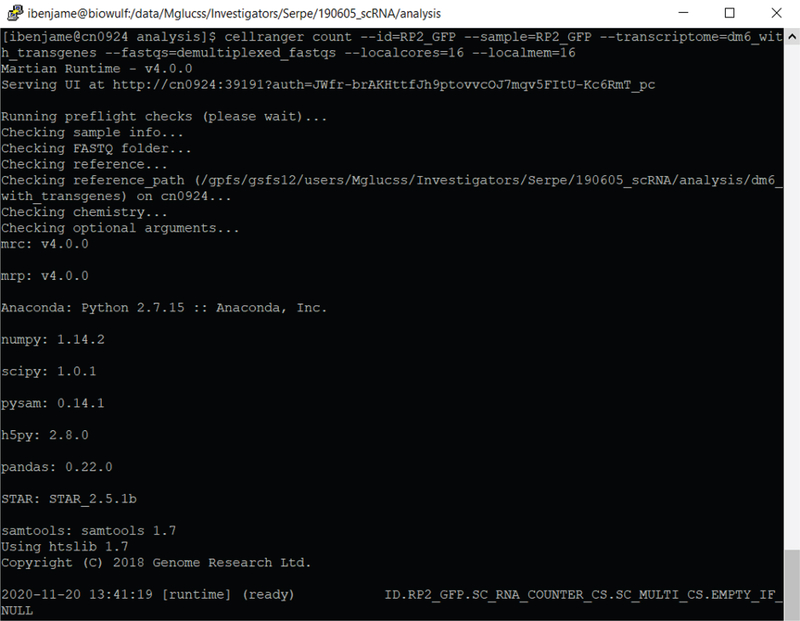
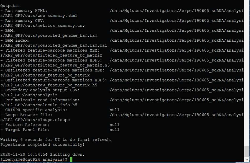
Drosophila provides a powerful genetic system and an excellent model to study the development and function of the nervous system. The fly's small brain and complex behavior has been instrumental in mapping neuronal circuits and elucidating the neural basis of behavior. The fast pace of fly development and the wealth of genetic tools has enabled systematic studies on cell differentiation and fate specification, and has uncovered strategies for axon guidance and targeting. The accessibility of neuronal structures and the ability to edit and manipulate gene expression in selective cells and/or synaptic compartments has revealed mechanisms for synapse assembly and neuronal connectivity. Recent advances in single-cell RNA sequencing (scRNA-seq) have further enhanced our appreciation and understanding of neuronal diversity in a fly brain. However, due to the small size of the fly brain and its constituent cells, scRNA-seq methodologies require a few adaptations. Here, we describe a set of protocols optimized for scRNA-seq analysis of the Drosophila larval ventral nerve cord, starting from tissue dissection and cell dissociation to cDNA library preparation, sequencing, and data analysis. We apply this workflow to three separate samples and detail the technical challenges associated with successful application of scRNA-seq to studies on neuronal diversity. An accompanying article (Vicidomini, Nguyen, Choudhury, Brody, & Serpe, 2021) presents a custom multistage analysis pipeline that integrates modules contained in different R packages to ensure high-flexibility, high-quality RNA-seq data analysis. These protocols are developed for Drosophila larval ventral nerve cord, but could easily be adapted to other tissues and model organisms. © 2021 U.S. Government. Basic Protocol 1: Dissection of larval ventral nerve cords and preparation of single-cell suspensions Basic Protocol 2: Preparation and sequencing of single-cell transcriptome libraries Basic Protocol 3: Alignment of raw sequencing data to indexed genome and generation of count matrices.

Keywords: 10× Genomics; Cell Ranger; Drosophila; cell dissociation; central nervous system; count matrix; demultiplexing; larval ventral nerve cord; reference genome; scRNA-seq.


Figures

Figure 1.
Figure 1.
Schematic of the Drosophila larval ventral nerve cord dissociation workflow Dissect wandering third instar Drosophila larvae, excise the VNCs and immediately immerse them in ice-cold Schneider’s medium supplemented with toxins (TTX, CNQX, AP-5) to inhibit neuronal firing. When all the VNCs are collected, initiate tissue dissociation by incubation with enzymes and follow up by mechanical disruption via trituration. Filter the dissociated cells to remove any remaining cluster, concentrate the cells and check their number and quality and viability.
Figure 2.
Figure 2.
Schematic of 10X Chromium platform Load the cell suspension and barcoded beads onto the 10X Chromium chip in lysis buffer. Cells and beads are combined into nanoliter-scale oil droplets to form an emulsion (called Gel bead-in emulsion). The single cell GEMs are immediately carried through reverse transcription reaction and PCR amplification. The resulting libraries are sequenced on an Illumina platform.
Figure 3.
Figure 3.
High sensitivity DNA analysis Bioanalyzer assay of full-length double stranded cDNAs (A) and sequencing libraries (B). Peaks at 35bp (red arrow) and 10380 bp (blue arrow) represent low- and high-molecular weight markers. The cDNAs isolated from the Drosophila third instar larvae VNCs have relatively high molecular weight, with a peak at ~700bp (black arrow). Upon PCR amplification, the libraries appear as a smooth peak.
Figure 4.
Figure 4.
Preparing the index genome “dm6_with_transgenes” with cellranger mkref
Figure 5.
Figure 5.
Diagram of the demultiplexing process with cellranger mkfastq for two samples/libraries sequenced together The base call files (BCLs) (binary files with raw scRNA-seq output generated by Illumina sequencers) and the sample sheet CSV files constitute the input for the cellranger mkfastq function. The output consists of three FASTQ files per sample; these are immediately subjected to quality control checks internally, by Cell Ranger suite algorithms. The FASTQ files are next used to identify and sort out the cells/barcodes, the unique molecular identifiers (UMIs) and the sample ID.
Figure 6.
Figure 6.
Example of syntax for running cellranger mkfastq
Figure 7.
Figure 7.
Example of cellranger count for sample “RP2_GFP” with built index “dm6_with_transgenes” deposited into folder “RP2_GFP”
Figure 8.
Figure 8.
Example of cellranger count outputs for sample “RP2_GFP” with built index “dm6_with_transgenes” deposited into folder “RP2_GFP”

Similar articles

Cited by

References

    1. Aicher TP, Carroll S, Raddi G, Gierahn T, Wadsworth MH 2nd, Hughes TK, Love C, and Shalek AK (2019). Seq-Well: A Sample-Efficient, Portable Picowell Platform for Massively Parallel Single-Cell RNA Sequencing. Methods Mol Biol 1979, 111–132. - PMC - PubMed
    1. Allen AM, Neville MC, Birtles S, Croset V, Treiber CD, Waddell S, and Goodwin SF (2020). A single-cell transcriptomic atlas of the adult Drosophila ventral nerve cord. Elife 9. - PMC - PubMed
    1. Alles J, Karaiskos N, Praktiknjo SD, Grosswendt S, Wahle P, Ruffault PL, Ayoub S, Schreyer L, Boltengagen A, Birchmeier C, et al. (2017). Cell fixation and preservation for droplet-based single-cell transcriptomics. BMC Biol 15, 44. - PMC - PubMed
    1. Bakken TE, Hodge RD, Miller JA, Yao Z, Nguyen TN, Aevermann B, Barkan E, Bertagnolli D, Casper T, Dee N, et al. (2018). Single-nucleus and single-cell transcriptomes compared in matched cortical cell types. PLoS One 13, e0209648. - PMC - PubMed
    1. Berck ME, Khandelwal A, Claus L, Hernandez-Nunez L, Si G, Tabone CJ, Li F, Truman JW, Fetter RD, Louis M, et al. (2016). The wiring diagram of a glomerular olfactory system. Elife 5. - PMC - PubMed

LinkOut - more resources