Review
. 2021 May;18(5):757-768.
doi: 10.1513/AnnalsATS.202006-696FR.

Excessive Daytime Sleepiness in Obstructive Sleep Apnea. Mechanisms and Clinical Management

Affiliations
Review

Excessive Daytime Sleepiness in Obstructive Sleep Apnea. Mechanisms and Clinical Management

Chitra Lal et al. Ann Am Thorac Soc. 2021 May.

Abstract

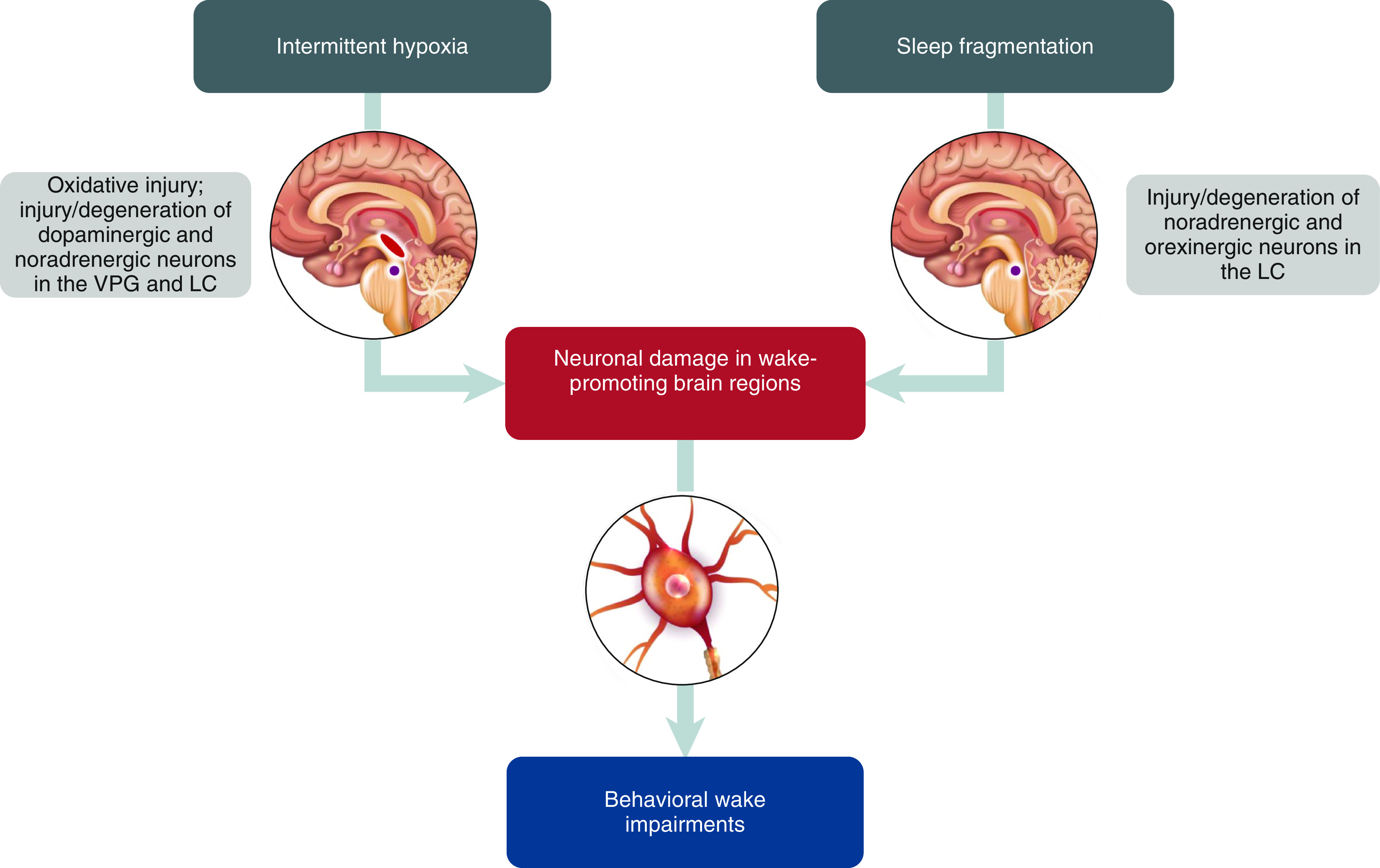
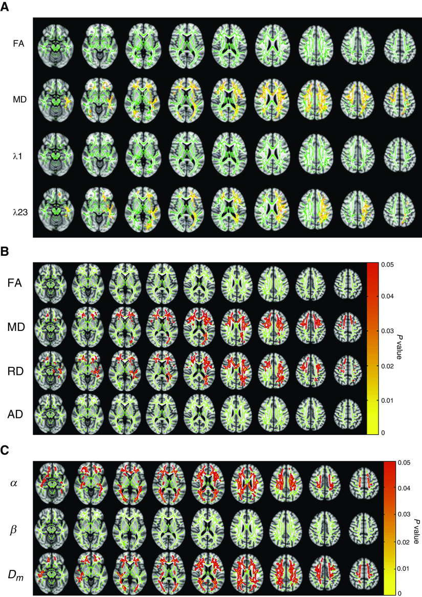
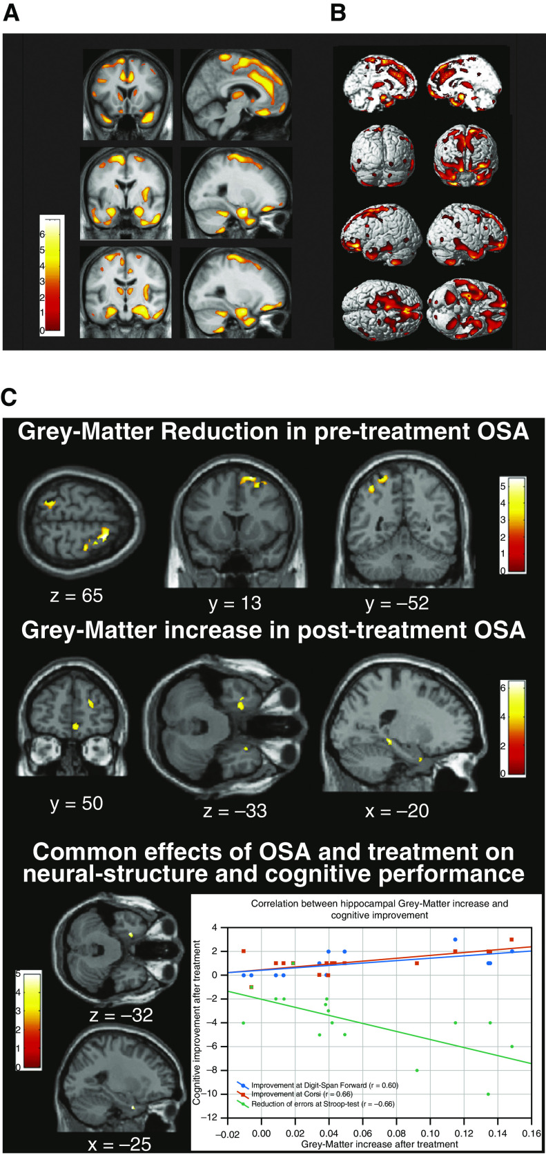
Many patients with obstructive sleep apnea (OSA) experience excessive daytime sleepiness (EDS), which can negatively affect daily functioning, cognition, mood, and other aspects of well-being. Although EDS can be reduced with primary OSA treatment, such as continuous positive airway pressure (CPAP) therapy, a significant proportion of patients continue to experience EDS despite receiving optimized therapy for OSA. This article reviews the pathophysiology and clinical evaluation and management of EDS in patients with OSA. The mechanisms underlying EDS in CPAP-treated patients remain unclear. Experimental risk factors include chronic intermittent hypoxia and sleep fragmentation, which lead to oxidative injury and changes in neurons and brain circuit connectedness involving noradrenergic and dopaminergic neurotransmission in wake-promoting regions of the brain. In addition, neuroimaging studies have shown alterations in the brain's white matter and gray matter in patients with OSA and EDS. Clinical management of EDS begins with ruling out other potential causes of EDS and evaluating its severity. Tools to evaluate EDS include objective and self-reported assessments of sleepiness, as well as cognitive assessments. Patients who experience residual EDS despite primary OSA therapy may benefit from wake-promoting pharmacotherapy. Agents that inhibit reuptake of dopamine or of dopamine and norepinephrine (modafinil/armodafinil and solriamfetol, respectively) have demonstrated efficacy in reducing EDS and improving quality of life in patients with OSA. Additional research is needed on the effects of wake-promoting treatments on cognition in these patients and to identify individual or disorder-specific responses.

Keywords: OSA; intermittent hypoxia; neurology; neuronal damage.


Figures

Figure 1.
Figure 1.
Proposed mechanisms of disease underlying residual excessive daytime sleepiness in obstructive sleep apnea. LC = locus coeruleus (purple); VPG = ventral periaqueductal gray (red).
Figure 2.
Figure 2.
White matter changes in patients with excessive daytime sleepiness associated with obstructive sleep apnea (OSA). (A) Diffusion tensor imaging (DTI) results showing possible white matter alterations in DTI metrics including FA, MD, λ1 (AD), and λ23 (RD) between sleepy and nonsleepy patients with OSA. Green: mean FA skeleton (threshold = 0.2) without significant change. Red-yellow: fibers with increased DTI metrics in the sleepy group when compared to the nonsleepy group (P < 0.05) (32). (B) Results from DTI whole-brain analysis based on FA, MD, λ23 (RD), and λ1 (AD) showing the presence or absence of differences between sleepy and nonsleepy patients with OSA. Age was included as a covariate in the group analyses for all parameters. Green: mean fractional anisotropy (FA) skeleton (threshold = 0.2) without significant change. Red-yellow: voxels with significantly increased parameters values in the sleepy group as compared to the nonsleepy group with P < 0.05 as shown in the color bar (33). (C) Whole-brain α, β, and Dm maps showing the presence or absence of differences between sleepy and nonsleepy patients with OSA (33). (A) Reprinted with permission of John Wiley & Sons. Xiong Y, et al. Brain white matter changes in CPAP-treated obstructive sleep apnea patients with residual sleepiness. J Magn Reson Imaging. 2017;45(5):1371–1378. Copyright © 2016 International Society for Magnetic Resonance in Medicine. (B and C) Reprinted from Zhang J, et al. White matter structural differences in OSA patients experiencing residual daytime sleepiness with high CPAP use: a non-Gaussian diffusion MRI study. Sleep Med. 2019;53:51–59. doi:10.1016/j.sleep.2018.09.011, with permission from Elsevier. AD = axial diffusivity; Dm = anomalous diffusion coefficient; FA = fractional anisotropy; MD = mean diffusivity; RD = radial diffusivity.
Figure 3.
Figure 3.
Gray matter changes in patients with OSA. (A) Areas of decreased gray matter concentration in patients with severe OSA (42). (B) Areas with reduced gray matter concentrations shown in three-dimensional rendering. Reduced gray matter concentrations were observed in the bilateral superior frontal gyri, left gyrus rectus, and bilateral frontomarginal gyri, bilateral anterior cingulate gyri, right anterior insular gyrus, bilateral caudate nuclei, bilateral thalami, bilateral amygdala and hippocampi, bilateral inferior temporal gyri, and bilateral cerebellar cortices. Results were superimposed on the two-dimensional planes of averaged T1 template of all subjects. Scales in color bar are t scores. Left-hand sides of images represent the left hemisphere of the brain. Reprinted with permission of Oxford University Press. Joo EY, et al. Reduced brain gray matter concentration in patients with obstructive sleep apnea syndrome. Sleep. 2010;33(2):235–241. (C) Gray matter volume before and after continuous positive airway pressure (CPAP) treatment in patients with OSA (46). Top row: Gray matter volume is decreased in untreated patients with OSA compared with control subjects. Middle row: Gray matter volume increased after CPAP treatment compared with before treatment. Bottom row: Gray matter in left hippocampal entorhinal cortex reduced before CPAP treatment and increased after treatment. There is also a correlation between the gray matter volume increase in this region and cognitive improvement after treatment. Reprinted by permission from Reference . OSA = obstructive sleep apnea.

References

    1. Kapur VK, Auckley DH, Chowdhuri S, Kuhlmann DC, Mehra R, Ramar K, et al. Clinical practice guideline for diagnostic testing for adult obstructive sleep apnea: an American Academy of Sleep Medicine clinical practice guideline. J Clin Sleep Med. 2017;13:479–504. - PMC - PubMed
    1. Benjafield AV, Ayas NT, Eastwood PR, Heinzer R, Ip MSM, Morrell MJ, et al. Estimation of the global prevalence and burden of obstructive sleep apnoea: a literature-based analysis. Lancet Respir Med. 2019;7:687–698. - PMC - PubMed
    1. American Academy of Sleep Medicine. International Classification of Sleep Disorders. 3rd ed. Darien, IL: American Academy of Sleep Medicine; 2014. Obstructive sleep apnea, adult; pp. 53–62.
    1. Sateia MJ. International classification of sleep disorders-third edition: highlights and modifications. Chest. 2014;146:1387–1394. - PubMed
    1. Pagel JF. Excessive daytime sleepiness. Am Fam Physician. 2009;79:391–396. - PubMed

Publication types

MeSH terms