. 2021 Feb 23;17(2):e1009331.
doi: 10.1371/journal.ppat.1009331. eCollection 2021 Feb.

Antibody affinity versus dengue morphology influences neutralization

Affiliations

Antibody affinity versus dengue morphology influences neutralization

Guntur Fibriansah et al. PLoS Pathog. .

Abstract

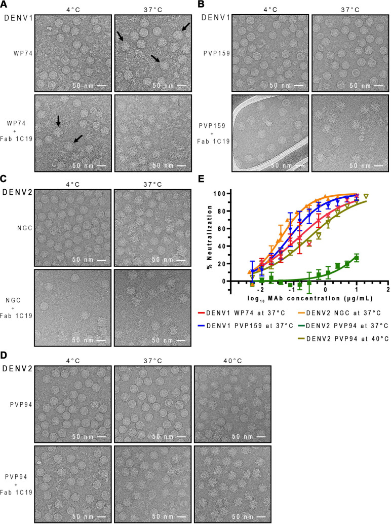
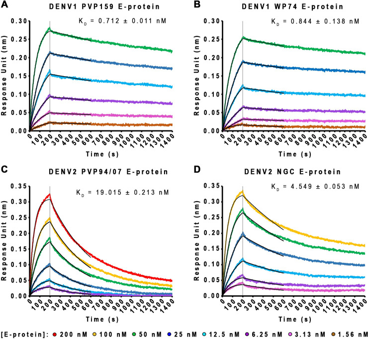
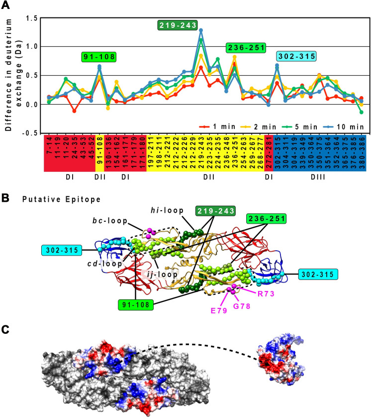
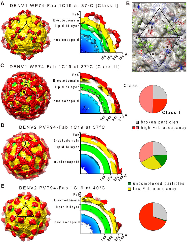
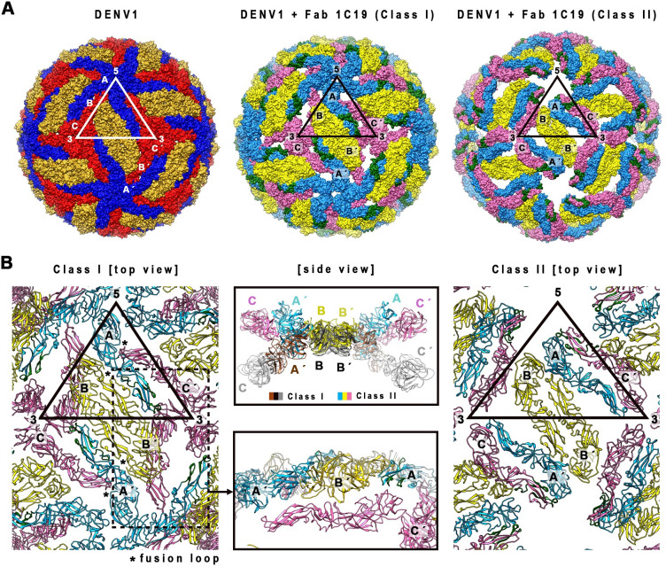
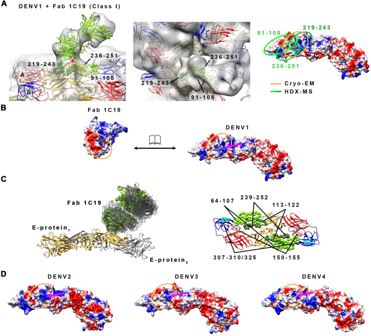
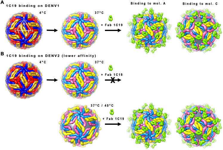
Different strains within a dengue serotype (DENV1-4) can have smooth, or "bumpy" surface morphologies with different antigenic characteristics at average body temperature (37°C). We determined the neutralizing properties of a serotype cross-reactive human monoclonal antibody (HMAb) 1C19 for strains with differing morphologies within the DENV1 and DENV2 serotypes. We mapped the 1C19 epitope to E protein domain II by hydrogen deuterium exchange mass spectrometry, cryoEM and molecular dynamics simulations, revealing that this epitope is likely partially hidden on the virus surface. We showed the antibody has high affinity for binding to recombinant DENV1 E proteins compared to those of DENV2, consistent with its strong neutralizing activities for all DENV1 strains tested regardless of their morphologies. This finding suggests that the antibody could out-compete E-to-E interaction for binding to its epitope. In contrast, for DENV2, HMAb 1C19 can only neutralize when the epitope becomes exposed on the bumpy-surfaced particle. Although HMAb 1C19 is not a suitable therapeutic candidate, this study with HMAb 1C19 shows the importance of choosing a high-affinity antibody that could neutralize diverse dengue virus morphologies for therapeutic purposes.


Conflict of interest statement

I have read the journal’s policy and the authors of this manuscript have the following competing interests: J.E.C. has served as a consultant for Lilly and Luna Biologics, is a member of the Scientific Advisory Boards of CompuVax and Meissa Vaccines and is Founder of IDBiologics. The Crowe laboratory at Vanderbilt University Medical Center has received sponsored research agreements from IDBiologics and AstraZeneca.

Figures

Fig 1
Fig 1. HMAb 1C19 binding and neutralization for DENV1 is independent of the virus morphologies, whereas for DENV2 the activities depend on virus changing to bumpy-surfaced morphology, which likely leads to improved epitope accessibility.
CryoEM micrographs showing binding of Fab 1C19 to (A) DENV1 WestPac 74, (B) DENV1 PVP159, (C) DENV2 NGC, (D) DENV2 PVP94. All upper rows show the morphology of un-complexed viruses at different temperatures and the lower rows show the 1C19 complexes. For DENV1 WestPac 74, the un-complexed virus that became bumpy-surfaced at 37°C are indicated by black arrows. For all viruses, Fab 1C19 did not bind well to virus at 4°C, suggesting that the epitope was partially hidden. One example is shown when Fab 1C19 was added to DENV1 WestPac 74 at 4°C, only some particles formed complexes with the Fab as indicated by black arrows in (A) bottom left panel. At 37°C, Fab 1C19 bound to both DENV1 strains well even though the un-complexed DENV1 strains had different morphologies- WestPac 74 showing 50% particles with bumpy surface and PVP159 remains smooth-surfaced. The Fab-complexed particles are spiky and appear larger than the bumpy un-complexed virus. For DENV2 strains, the un-complexed NGC and PVP94 have a bumpy or smooth surface morphology, respectively. It appears that Fab 1C19 binds well to the bumpy-surfaced strain NGC, but poorly to the strain PVP94, as observed by 50% spiky particles in the PVP94 + Fab 1C19 sample. Increasing the temperature to 40°C increased the number of bumpy-surfaced particles in the un-complexed DENV2 PVP94 control leading to complete binding of Fab 1C19. (E) Neutralization profile of HMAb 1C19 for DENV1 or DENV2 strains. HMAb 1C19 neutralized DENV1 strains WestPac 74 and PVP159 equally well, whereas for DENV2, the antibody neutralized only strain NGC but not PVP94/07. HMAb 1C19 neutralized DENV2 strain PVP94/07 only at 40°C. The experiment was repeated three times. The neutralization profiles correlated well with the binding properties of Fab 1C19 observed in cryoEM.
Fig 2
Fig 2. HMAb 1C19 showed higher affinity of binding to E protein of DENV1 than to that of DENV2.
(A-D) Bio-layer interferometry curves (in different colors) and fitting curves (in black) for binding of increasing concentrations of DENV1 or DENV2 rE protein to immobilized IgG 1C19. These results showed HMAb 1C19 has a higher affinity of binding to DENV1 than to DENV2 E protein.
Fig 3
Fig 3. HDXMS experiments showed that the 1C19 epitope contains residues in peptides 91–108, 219–243 and 236–251 on DII.
(A) Plot of the deuterium exchange differences between pepsin-generated peptide fragments of the un-complexed rE protein and the corresponding peptides from rE protein-Fab 1C19 complex at different incubation timings 1 (red line), 2 (yellow line), 5 (green line) or 10 (blue line) mins. Higher deuterium exchange difference indicates that the peptide has lower solvent accessibility when bound by Fab 1C19. Each dot on the plot represents a peptide fragment of the rE protein, as indicated on the x-axis. The domain where the peptide, on the x-axis, resides on the E protein is indicated by its box color: red, yellow or blue for DI, DII or DIII, respectively. Difference in deuterium exchange (deuterium alterations) by > 1 Da was considered significant, whereas between 0.5–1 Da was moderate. The peptides that showed moderate alterations may represent sites on the E protein that had weak binding or had undergone distant conformational changes upon antibody binding. Based on their deuterium alterations, peptides can be assigned into three groups. The first group is for peptides that showed significant alteration in deuterium exchange (peptide 219–243, dark green filled box). The second group consists of peptides with the second highest deuterium alteration, peptides 91–108 and 236–251, light green filled box. The third group contains other peptides with moderate deuterium alterations (peptide 302–315, cyan filled box). (B) Location of the peptides with high or moderate deuterium alterations on the E protein structure. The putative epitope consisting of residues in peptides 91–108 (light green), 219–243 (dark green) or 236–251 (light green) are shown as spheres on the rE protein structure. Overlapping residues between peptides 219–243 and 236–251 are colored as olive-green spheres. DI, II or III of E protein are colored in red, yellow or blue, respectively. Residues that had been shown by site-directed mutagenesis studies to be important for HMAb 1C19 binding [33], are shown as magenta spheres. Peptide 302–315 is shown as cyan spheres. (C) The surface charges of peptides 91–108 (c strand and cd loop), 219–243 (hi loop), 236–251 (ij loop) in an E protein dimer are shown on the left, while the variable region of Fab 1C19 is on the right. Positive, neutral or negative charges are shown in blue, white or red colors, respectively. Charges of the putative epitope on the E protein and Fab seemed to be complementary to each other (arrow dashed line).
Fig 4
Fig 4. The cryo-EM density maps of DENV1 strain WestPac 74:Fab 1C19 complex.
(A) Class I or (C) Class II particles at 37°C, and DENV2 strain PVP94/07:Fab 1C19 complex at (D) 37°C or (E) 40°C. For all cryoEM maps, the surface (left) and center cross-section (center) are shown. (A) The cryo-EM map of Class I DENV1 strain WestPac 74:Fab 1C19 complex particles showed 60 copies of Fab (red) bound to the virus surface (yellow). The black triangle represents an icosahedral asymmetric unit, with the 2-, 3- and 5-fold vertices indicated. (B) The fit of E proteins and Fab 1C19 (light green) molecules into the Class I DENV1 strain WestPac 74:Fab 1C19 at 37°C cryo-EM map. All E protein and Fab molecule models were fitted into density, none were in negative densities and the molecules did not clash with each other. Three individual E protein molecules in an asymmetric unit are labelled as A, B and C, whereas the corresponding molecules in neighboring asymmetric unit within a raft are labelled as A′, B′ and C′. DI, II and III of E protein are colored in red, yellow and blue, respectively. (C) The Class II DENV1 strain WestPac 74:Fab 1C19 at 37°C map likely has 60 copies of Fab molecules bound (black arrow). (D) Cryo-EM map of DENV2 strain PVP94/07:Fab 1C19 complex at 37°C showed similarity with Class II DENV1 strain WestPac 74:Fab 1C19 complex at 37°C, whereas the complex at (E) 40°C appeared to be similar to Class I DENV1 strain WestPac 74:Fab 1C19 complex at 37°C. In the rightmost panel of (C), (D) and (E), the proportion of the different particles within the virus population (right) is shown as a pie chart.
Fig 5
Fig 5. E protein quaternary structure changes in the DENV1 strain WestPac 74-Fab 1C19 Class I and II structures compared to the un-complexed compact smooth DENV1 structure.
(A) E protein molecules organization on the un-complexed DENV1 (left) and the DENV1-Fab 1C19 complex Class I (middle) and II structures (right). The E protein molecules are shown as surface representation. In the un-complexed compacted smooth surfaced DENV1 structure, mols A, B or C E proteins are colored in blue, gold, or red, respectively, whereas the same molecules of the DENV1-Fab 1C19 complexes are in lighter shades of the same color (light blue, yellow or pink). The putative 1C19 epitope identified by HDXMS on mol A or C in the Class I and Class II structures, respectively are colored in dark green. (B) The E protein arrangements in Class I (left) and II (right) of DENV1-Fab 1C19 complex structures. (Left) E protein arrangement in Class I structure. Mol C (pink) in the complex structure did not clash with the adjacent molecules and is located on a lower radius. (Center bottom), Side view of the zoomed-in area of the dotted box on the left. (Center top) Superposition of an E-protein raft of Class I structure onto that of Class II structure. The E protein mols A and C of Class II complex particles are located at higher radii than those of Class I complex particles, while the E protein dimer mols B-B′ is at the same radius.
Fig 6
Fig 6. The likely interactions between Fab 1C19 and DENV1 E protein, as suggested by combining HDXMS results, the cryo-EM structure and MD simulations, and its comparison to the other DENV serotypes.
(A) (Left) Zoom-in side view of the fit of Fab 1C19 molecules (light green) and E proteins in the cryo-EM map (gray transparent) of Class I particles. The putative epitope identified by HDXMS consists of residues in peptides 91–108, 219–243 and 236–251 (Fig 3B) are indicated by green spheres of different shades. The glycosylation at residue N67 is indicated by red star. (Center) Top view of Fab 1C19 density on fitted E protein. Fab 1C19 densities are located right on top of the putative epitope identified by HDXMS, mainly on peptides 219–243 and 236–251. (Right) The HDXMS results helped in guiding the fit of the molecule into the cryo-EM map, thus identifying the location of the 1C19 epitope on the E protein. The epitope size identified by HDXMS (light and dark green lines) is larger than that identified cryo-EM (orange circle). (B) The electrostatic charges on the interface surfaces of Fab 1C19 and DENV1 E protein (open book representation), identified by combining both techniques, are complementary to each other. Blue, white or red colors indicate positive, neutral or negative charges, respectively. The dashed orange lines indicate the border of heavy and light chains on the antibody paratope (left) and the corresponding footprint on the E protein (right). The magenta arrow indicates the positively charge patch on E protein that likely interacts with a negatively charged patch on the antibody paratope. (C) MD simulations reveal the highly dynamic properties of Fab 1C19 and its epitope on DENV1 E protein. (Left) Side view of E protein—Fab 1C19 complex. E protein dimer is shown in cartoon representation (grey and gold yellow colors) together with Fab 1C19 in its initial (green) near-experimental conformation. All visited states of C19 Fab over the entire simulation time are shown in transparent grey cartoon representation. (Right) The top view of E protein dimer shown in cartoon representation (domain I: red, domain II: yellow, domain III: blue) with the three distinctive regions (shown as spheres). (D) The electrostatic potential surface of the epitope on the other DENV serotypes. The epitope on DENV2 and DENV3 has a similar positively-charged surface (magenta arrow) as that on DENV1, while the same region on DENV4 is the least positively charged surface. This finding is consistent with the neutralization profile of HMAb 1C19, which showed potent neutralization of viruses of the DENV1-3 serotype but not the DENV4 serotype. DENV structures used for electrostatic potential surface analyses were the cryo-EM structures of DENV1, DENV2, DENV3 or DENV4 (PDB codes 4CCT, 3J27, 3J6S or 4CBF, respectively).
Fig 7
Fig 7. Binding of Fab 1C19 to DENV1 and DENV2.
Fab 1C19 binds to a partially hidden epitope on DII of the E proteins. It has a strong affinity to DENV1 E protein and therefore it can out-compete E-to-E interactions even when the virus maintains a smooth compact surface that only partially reveals the epitope. Fab 1C19 can only bind to one of the E proteins of the A-C dimer, depending on which is exposed first when the E protein is vibrating at 37°C. Fab 1C19 binds in a similar same way to DENV2 (either mol A or C), but because its affinity to DENV2 epitope is lower, the virus has to change its conformation to a looser “bumpy” surface structure at higher temperatures (37°C or 40°C) to expose its epitope fully before the antibody can bind.

References

    1. Bhatt S, Gething PW, Brady OJ, Messina JP, Farlow AW, Moyes CL, et al.. The global distribution and burden of dengue. Nature. 2013;496(7446):504–7. 10.1038/nature12060 - DOI - PMC - PubMed
    1. Villar L, Dayan GH, Arredondo-Garcia JL, Rivera DM, Cunha R, Deseda C, et al.. Efficacy of a tetravalent dengue vaccine in children in Latin America. The New England journal of medicine. 2015;372(2):113–23. 10.1056/NEJMoa1411037 . - DOI - PubMed
    1. Capeding MR, Tran NH, Hadinegoro SR, Ismail HI, Chotpitayasunondh T, Chua MN, et al.. Clinical efficacy and safety of a novel tetravalent dengue vaccine in healthy children in Asia: a phase 3, randomised, observer-masked, placebo-controlled trial. Lancet. 2014;384(9951):1358–65. 10.1016/S0140-6736(14)61060-6 . - DOI - PubMed
    1. Guy B, Briand O, Lang J, Saville M, Jackson N. Development of the Sanofi Pasteur tetravalent dengue vaccine: One more step forward. Vaccine 2015. 10.1016/j.vaccine.2015.09.108 . - DOI - PubMed
    1. Sabchareon A, Wallace D, Sirivichayakul C, Limkittikul K, Chanthavanich P, Suvannadabba S, et al.. Protective efficacy of the recombinant, live-attenuated, CYD tetravalent dengue vaccine in Thai schoolchildren: a randomised, controlled phase 2b trial. The Lancet. 2012;380(9853):1559–67. 10.1016/S0140-6736(12)61428-7 - DOI - PubMed

Publication types

MeSH terms