Review
. 2021 Apr:20:139-162.
doi: 10.1016/j.jtos.2021.02.007. Epub 2021 Feb 20.

The ocular surface immune system through the eyes of aging

Affiliations
Review

The ocular surface immune system through the eyes of aging

Jeremias G Galletti et al. Ocul Surf. 2021 Apr.

Abstract

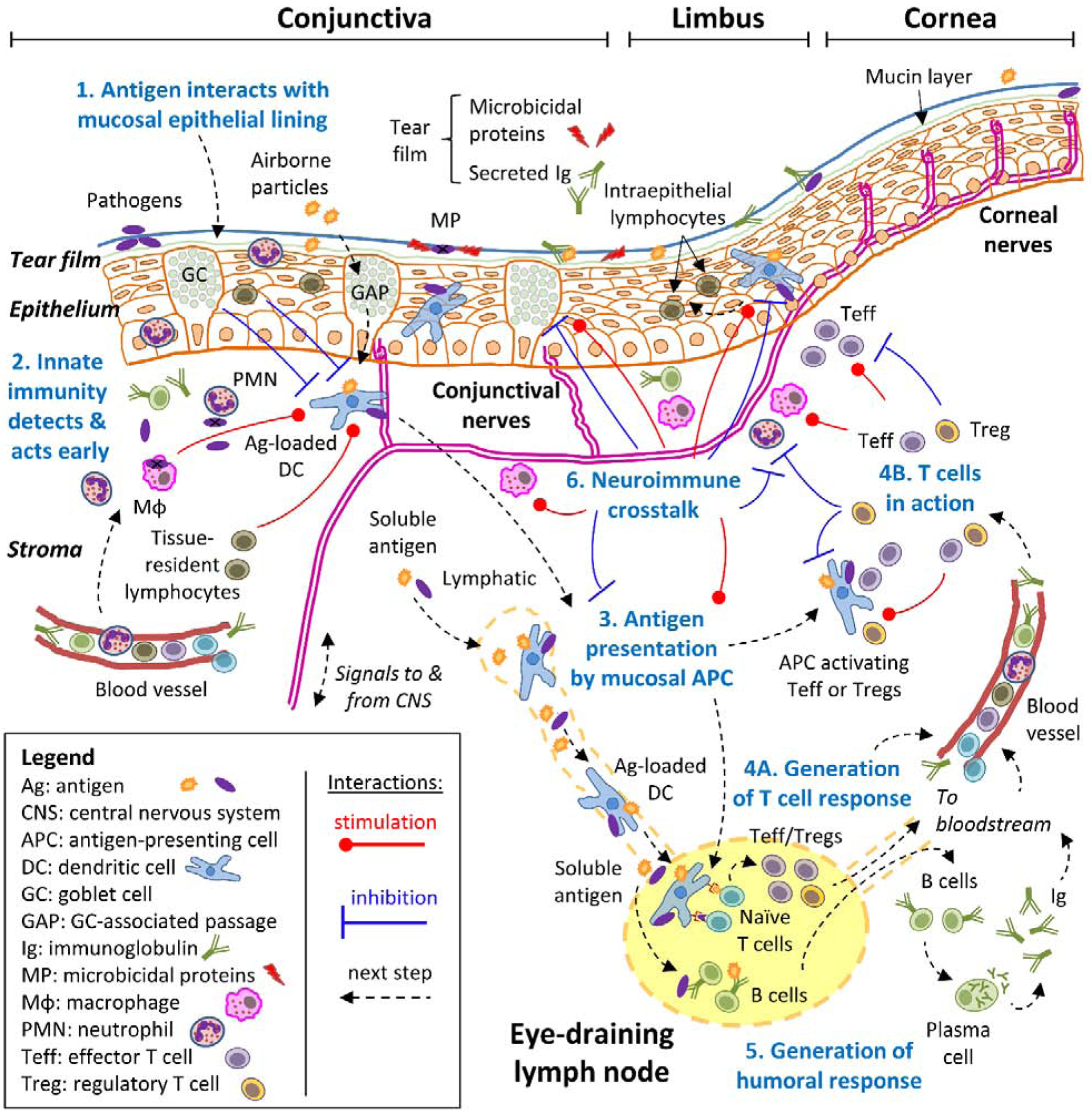
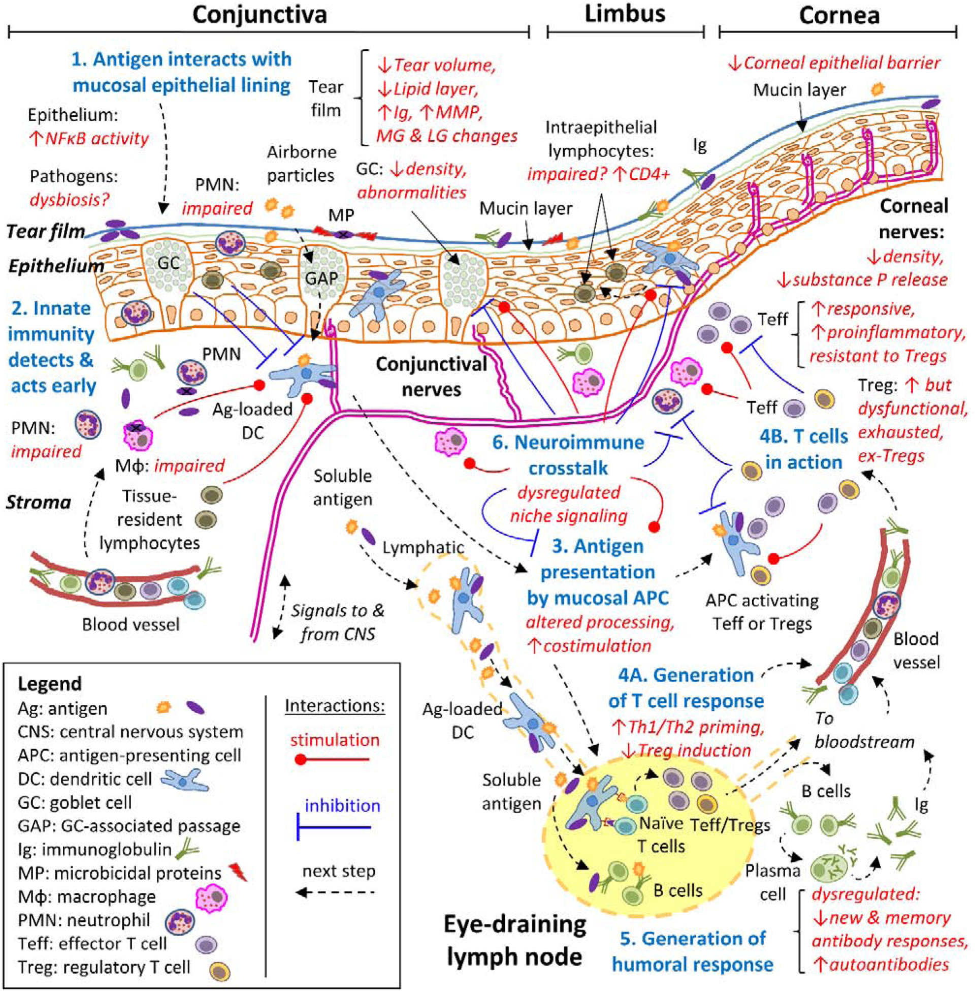
Since the last century, advances in healthcare, housing, and education have led to an increase in life expectancy. Longevity is accompanied by a higher prevalence of age-related diseases, such as cancer, autoimmunity, diabetes, and infection, and part of this increase in disease incidence relates to the significant changes that aging brings about in the immune system. The eye is not spared by aging either, presenting with age-related disorders of its own, and interestingly, many of these diseases have immune pathophysiology. Being delicate organs that must be exposed to the environment in order to capture light, the eyes are endowed with a mucosal environment that protects them, the so-called ocular surface. As in other mucosal sites, immune responses at the ocular surface need to be swift and potent to eliminate threats but are at the same time tightly controlled to prevent excessive inflammation and bystander damage. This review will detail how aging affects the mucosal immune response of the ocular surface as a whole and how this process relates to the higher incidence of ocular surface disease in the elderly.

Keywords: Aging; Goblet cells; Immune cells; Lacrimal gland; T regulatory t cells; Tear film.


Conflict of interest statement

Disclosure: No financial interests to disclose.

Figures

Figure 1:
Figure 1:. Schematic mucosal immune response of the ocular surface.
A simplified approach to the mucosal immune response to diverse antigens, consisting of 6 consecutive steps (numbered in blue), is exemplified and adopted throughout this review. For clarity, all steps revolve around the concept of an antigen (the molecule or molecular structure against which the adaptive immune system reacts), which can derive from pathogens, foreign substances, or even self-tissues. Step 1: the antigen interacts with the mucosal epithelial lining, which acts as a barrier aided by blinking action and the tears. Step 2: antigens may breach through the epithelial barrier, setting off the innate immune system’s detection systems because receptors on diverse cell types recognize molecular patterns that are associated with potentially dangerous entities. This triggers a swift defense that results in early containment and that influences the next step at the same time. Step 3: specialized antigen-presenting cells (APC) that reside in the ocular surface capture antigens and migrate to the eye-draining lymph nodes, where they process them and present antigen-derived peptides on their surface. Step 4: naïve T cells that continuously recirculate through lymph nodes recognize specific peptides on APC, become activated, and then expand and differentiate to either effector (Teff) or regulatory (Treg) T cells depending on additional signals that they pick up during antigen presentation. These antigen-experienced T cells leave the lymph node and through the bloodstream, reach the ocular surface where they can exert their many functions in the immune response. Step 5: concomitantly with step 3 and 4, soluble antigens reach the lymph node from the ocular surface, where B cells might interact with them, become activated, and then expand and differentiate into plasma cells that migrate to other tissues through the bloodstream, mainly the bone marrow. Once at their final destination, plasma cells secrete large amounts of immunoglobulin (Ig) that reach the ocular surface and the lacrimal glands through the bloodstream. Step 6: the nervous system can also sense signals associated with some antigens and interacts with every cell type of the immune response in a bidirectional process (neuroimmune regulation). Although most of the immune response takes place at the ocular surface, it should be noted that critical steps 3, 4, and 5 begin in the draining lymph node. Also, the immune system is continuously interacting with various antigens, and thus, these processes take place simultaneously.
Figure. 2.
Figure. 2.. Tear film and lacrimal gland changes during aging.
A. Tear washings from young and aged mice of both sexes were collected, and immunoglobulins were measured using Luminex assay. Each dot corresponds to tear washings from 10 animals (20 eyes). B. Representative macro images of young and aged female lacrimal glands. Note the yellowish-tan appearance of aged lacrimal gland and presence of cysts (present in 20–30% of aged lacrimal glands, F2 and F3). C. Representative scans of lacrimal gland sections stained with H&E from young and aged B6 female mice. Areas of lymphocytic infiltration are demarcated aged glands (F1), and areas of dilated ducts/cysts are easily identified (F2, asterisks). 2M = 2 months; F = female.
Figure 3:
Figure 3:. Ocular surface changes during aging.
A. Representative laser scanning confocal microscope images of whole mounts of cornea and conjunctiva stained with occludin (green) and Hoechst 33342 DNA staining (blue). Arrows indicate desquamating cells. B. Representative bright-field images of conjunctiva sections showing PAS+ cells (purple-magenta). Insets are high magnification of a demarcated area. C. Representative laser scanning confocal microscope images of conjunctiva cryosections stained with MHC II (green), CD11c (red) and CD11b (white), and Hoechst 33342 nuclear staining (blue). Asterisks indicate goblet cells; the dotted area is magnified on the right. D. Representative whole-mount images of corneas stained with β-III tubulin antibody ([421], published under a Creative Commons CC BY 4.0 license). 24M = 24 months
Figure 4:
Figure 4:. Schematic mucosal immune response of the aged ocular surface.
Summary of the most relevant age-related changes presented in this review. As for figure 1, a simplified, 6-step approach to the mucosal immune response to diverse antigens is shown in the context of aging. Age-related changes are described in red italics, affecting steps 1 (antigen interaction with the mucosal lining), 2 (the innate immune response), 3 (antigen presentation), 4 (generation of a T cell response), 5 (generation of a humoral response), and 6 (neuroimmune interactions). See Figure 1 for a more detailed description of each step.

References

    1. Stern ME, Beuerman RW, Fox RI, Gao J, Mircheff AK, Pflugfelder SC. A unified theory of the role of the ocular surface in dry eye. AdvExpMedBiol. 1998;438:643–51. - PubMed
    1. Cher I Ocular surface concepts: Development and citation. Ocular Surface. 2014;12:10–3. - PubMed
    1. Knop E, Knop N. The role of eye-associated lymphoid tissue in corneal immune protection. JAnat. 2005;206:271–85. - PMC - PubMed
    1. Galletti JG, Guzman M, Giordano MN. Mucosal immune tolerance at the ocular surface in health and disease. Immunology. 2017;150:397–407. - PMC - PubMed
    1. Foulsham W, Coco G, Amouzegar A, Chauhan SK, Dana R. When Clarity Is Crucial: Regulating Ocular Surface Immunity. Trends in immunology. 2018;39:288–301. - PMC - PubMed

Publication types