. 2021 Feb;11(2):200409.
doi: 10.1098/rsob.200409. Epub 2021 Feb 24.

Ectopic positioning of the cell division plane is associated with single amino acid substitutions in the FtsZ-recruiting SsgB in Streptomyces

Affiliations

Ectopic positioning of the cell division plane is associated with single amino acid substitutions in the FtsZ-recruiting SsgB in Streptomyces

Xiansha Xiao et al. Open Biol. 2021 Feb.

Abstract

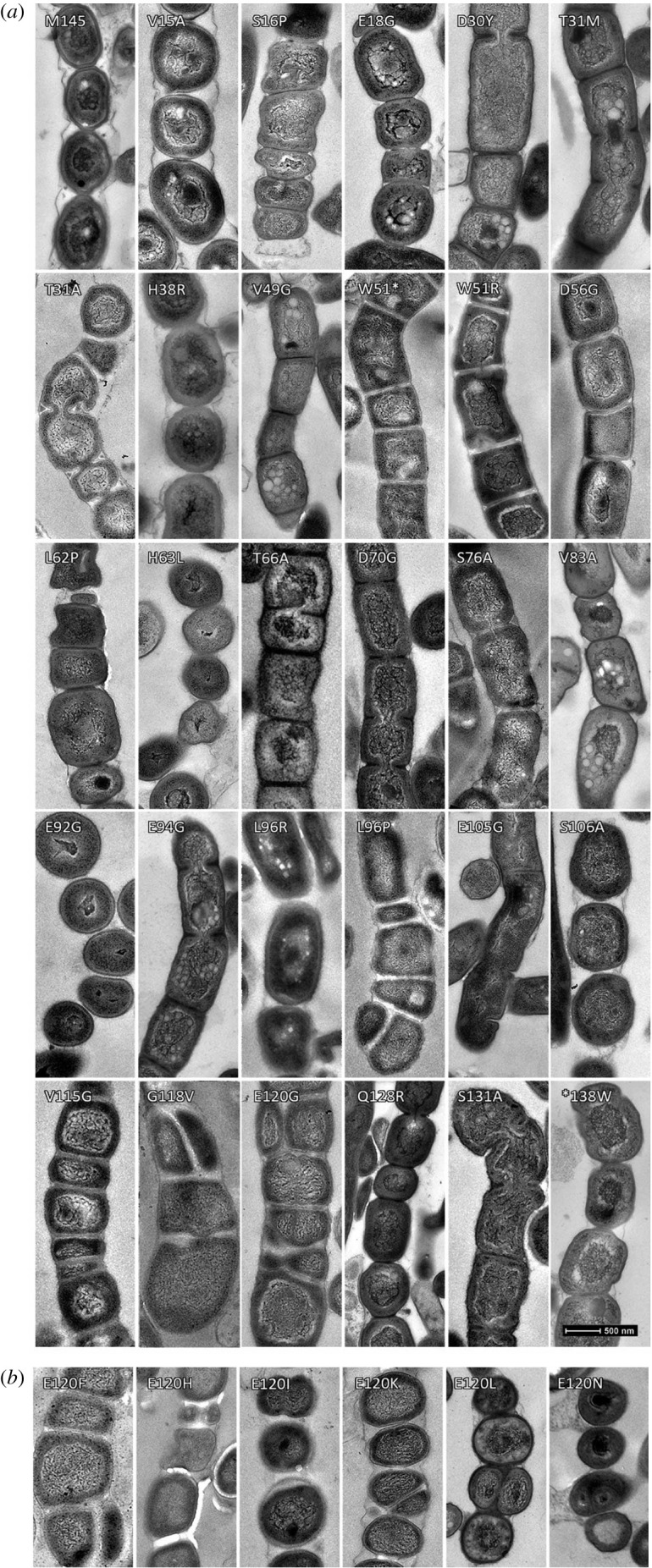
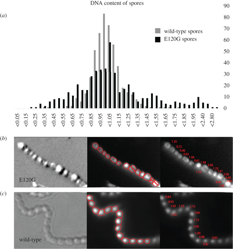
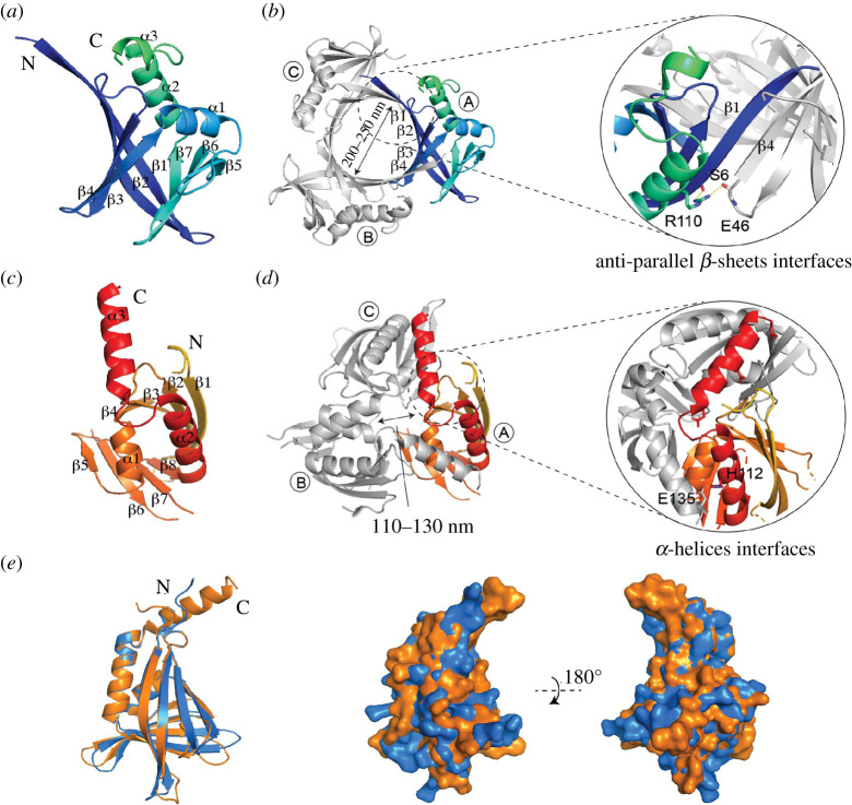
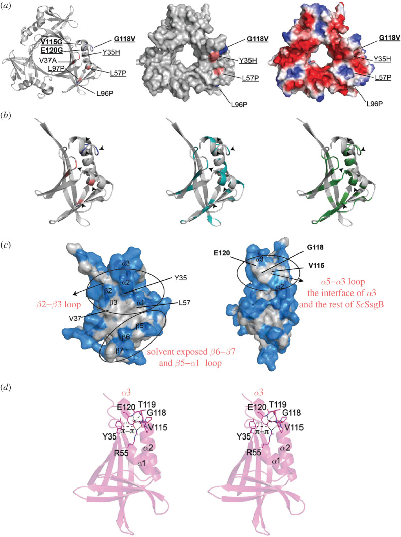
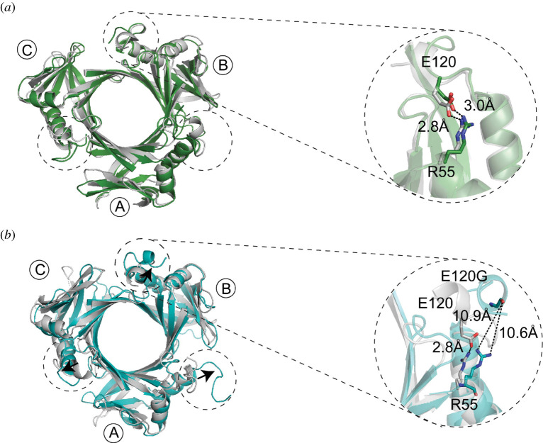
In most bacteria, cell division begins with the polymerization of the GTPase FtsZ at mid-cell, which recruits the division machinery to initiate cell constriction. In the filamentous bacterium Streptomyces, cell division is positively controlled by SsgB, which recruits FtsZ to the future septum sites and promotes Z-ring formation. Here, we show that various amino acid (aa) substitutions in the highly conserved SsgB protein result in ectopically placed septa that sever spores diagonally or along the long axis, perpendicular to the division plane. Fluorescence microscopy revealed that between 3.3% and 9.8% of the spores of strains expressing SsgB E120 variants were severed ectopically. Biochemical analysis of SsgB variant E120G revealed that its interaction with FtsZ had been maintained. The crystal structure of Streptomyces coelicolor SsgB was resolved and the key residues were mapped on the structure. Notably, residue substitutions (V115G, G118V, E120G) that are associated with septum misplacement localize in the α2-α3 loop region that links the final helix and the rest of the protein. Structural analyses and molecular simulation revealed that these residues are essential for maintaining the proper angle of helix α3. Our data suggest that besides altering FtsZ, aa substitutions in the FtsZ-recruiting protein SsgB also lead to diagonally or longitudinally divided cells in Streptomyces.

Keywords: SsgB; cell division; crystal structure; ectopic septa; mutagenesis.


Figures

Figure 1.
Figure 1.
Transmission electron micrographs of sporogenic hyphae from the wild-type and single sporulating SsgB mutants. (a) D30Y, V49G, T66A, D70G, E94G and S131A result in thinner cell walls. L96P, V115G, G118V and E120G give rise to septum rotation, which also affect DNA condensation. Disturbed DNA segregation was observed in L88R (see electronic supplementary material, figure S4). Mutants expressing SsgB variants V15A, S16P, E18G, T31A, T31M, H38R, W51R, L62P, H63L, S76A, V83A, E92G, L96R, E105G, S106A and Q128R showed aberrant spore sizes (see also electronic supplementary material, table S2). (b) Six additional amino acid substitutions of E120 that lead to longitudinal division. (E120G is shown in figure 1a). Bars: 500 nm for TEM micrographs.
Figure 2.
Figure 2.
Impression prints of spores from cells expressing wild-type SsgB or SsgB E120 mutants. (a) Longitudinal cell division (pointed by arrowhead) that are shown by fluorescent microscope from different E120 mutants. Left, bright-field images; middle, Syto9/PI stained images; right, overlays of the two images. All the spores were obtained after 7 days of growth from wild-type cells or from transformants of its ssgB null mutant expressing SsgB E120 mutants. Bars, 1 µm. (b) SEM imaging of wild-type SsgB and its E120G mutant. Panel (ii) shows the variability in spore size, whereas panels (iii) and (iv) show the start of longitudinal indentations and a later stage of longitudinal division, respectively.
Figure 3.
Figure 3.
Intensity plots of SsgB foci on the septa. (a) The wild-type SsgB-localization during early cell division shows two foci on either side of the hyphal wall; (b) Mutants expressing SsgB (E120G) showed aberrant localization, whereby SsgB was located all over the hyphal wall; (c) Occasional longitudinal septation was seen in SsgB (G118V), whereby eGFP fusions of SsgB mutant proteins localized parallel to the hyphal wall and in the middle of the hyphae. The red box indicates the width of the box that was used for making the profiles. Y1–Y5, single intensity plots of selected foci; X-axis, the distance between two foci on either side of hyphal wall; Y-axis, the fluorescent intensity.
Figure 4.
Figure 4.
DNA content analysis of SsgB variants. (a) The DNA content distribution in both wild-type (light grey) and E120G spores (black) is shown. The median DNA content of the spores was set to 1 to normalize the data. A normal distribution was observed for wild-type strain, whereas the strain expressing SsgB (E120G) had much more variation in DNA content. Y-axis, the frequency at which each DNA content was observed; X-axis, the DNA content in each spore. (b) To illustrate, one spore chain containing longitudinal divisions is shown with the respective DNA content in each spore (between 0.4 and 3.0 chromosomes). (c) In comparison, wild-type spores showed relatively little variation (between 0.83 and 1.15 in the spore chain).
Figure 5.
Figure 5.
Crystal structure of the ScSsgB trimer. (a) Ribbon diagrams showing the monomer structure of SsgB from S. coelicolor. (b) The overall structure of ScSsgB reveals a trimer. Structure statistics are listed in electronic supplementary material, table S6. The interface between adjacent monomers is formed by two antiparallel β-sheets. (c) The monomer structure of SsgB from T. fusca (PDB code 3CM1). (d) The interface between adjacent monomers of Tf SsgB is formed by α-helices. (e) Overlap of ScSsgB (blue) and Tf SsgB (orange) subunits. Left, side view of the electrostatic surface alignment of ScSsgB and Tf SsgB structure. Right, the same electrostatic figure but rotated by 180°.
Figure 6.
Figure 6.
Key mutations and their interactions in SsgB trimer structure. (a) Left, aa substitutions L96P, V115G, G118V and E120G show tilted division; aa substitutions of Y35H, V37A, L57P and L97P (deep salmon) result in non-sporulating phenotype; middle, right, top view of all the functional aa substitutions mapped on the surface structures. Conserved residues are underlined. (b) Left, key residues as shown by (a); middle, residues corresponding to all substitutions in this study (cyan); right, conserved residues (green). (c) Key residues (Y35H, V37A, L57P and L97P) are located in the hydrophobic patch, while V115, G118 and E120 are clustered on the α2–α3 loop region that mediates the interactions of α3 and the globular domain of SsgB. White patches in the surface structure indicate hydrophobic residues. (d) Stereo-view of E120 in the monomer structure and its interactions with the surrounding residues.
Figure 7.
Figure 7.
Molecular dynamic simulation of SsgB wild-type and E120G mutant. (a) MD result of SsgB wild-type structure. The α3 helix stays at the same orientation and the distance between R55 and E120 keeps at 2.8 Å and 3.0 Å, before (green) and after MD (grey). (b) The arrows indicate the changed angle of SsgB-E120G mutant (cyan) compared to the wild-type SsgB (grey) after MD.

Similar articles

Cited by

References

    1. Bi EF, Lutkenhaus J. 1991. FtsZ ring structure associated with division in Escherichia coli. Nature 354, 161-164. (10.1038/354161a0) - DOI - PubMed
    1. Mukherjee A, Lutkenhaus J. 1994. Guanine nucleotide-dependent assembly of FtsZ into filaments. J. Bacteriol. 176, 2754-2758. (10.1128/JB.176.9.2754-2758.1994) - DOI - PMC - PubMed
    1. Erickson HP, Taylor DW, Taylor KA, Bramhill D. 1996. Bacterial cell division protein FtsZ assembles into protofilament sheets and minirings, structural homologs of tubulin polymers. Proc. Natl Acad. Sci. USA 93, 519-523. (10.1073/pnas.93.1.519) - DOI - PMC - PubMed
    1. de Boer P, Crossley R, Rothfield L. 1992. The essential bacterial cell-division protein FtsZ is a GTPase. Nature 359, 254-256. (10.1038/359254a0) - DOI - PubMed
    1. Lyu Z, Coltharp C, Yang X, Xiao J. 2016. Influence of FtsZ GTPase activity and concentration on nanoscale Z-ring structure in vivo revealed by three-dimensional Superresolution imaging. Biopolymers 105, 725-734. (10.1002/bip.22895) - DOI - PMC - PubMed

Publication types

Substances

LinkOut - more resources