Review
. 2021 Feb;11(2):200359.
doi: 10.1098/rsob.200359. Epub 2021 Feb 24.

The origin of animals: an ancestral reconstruction of the unicellular-to-multicellular transition

Affiliations
Review

The origin of animals: an ancestral reconstruction of the unicellular-to-multicellular transition

Núria Ros-Rocher et al. Open Biol. 2021 Feb.

Abstract

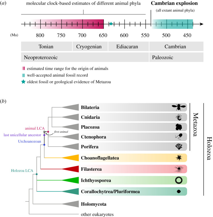
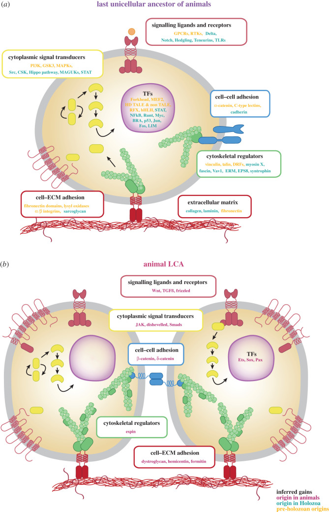
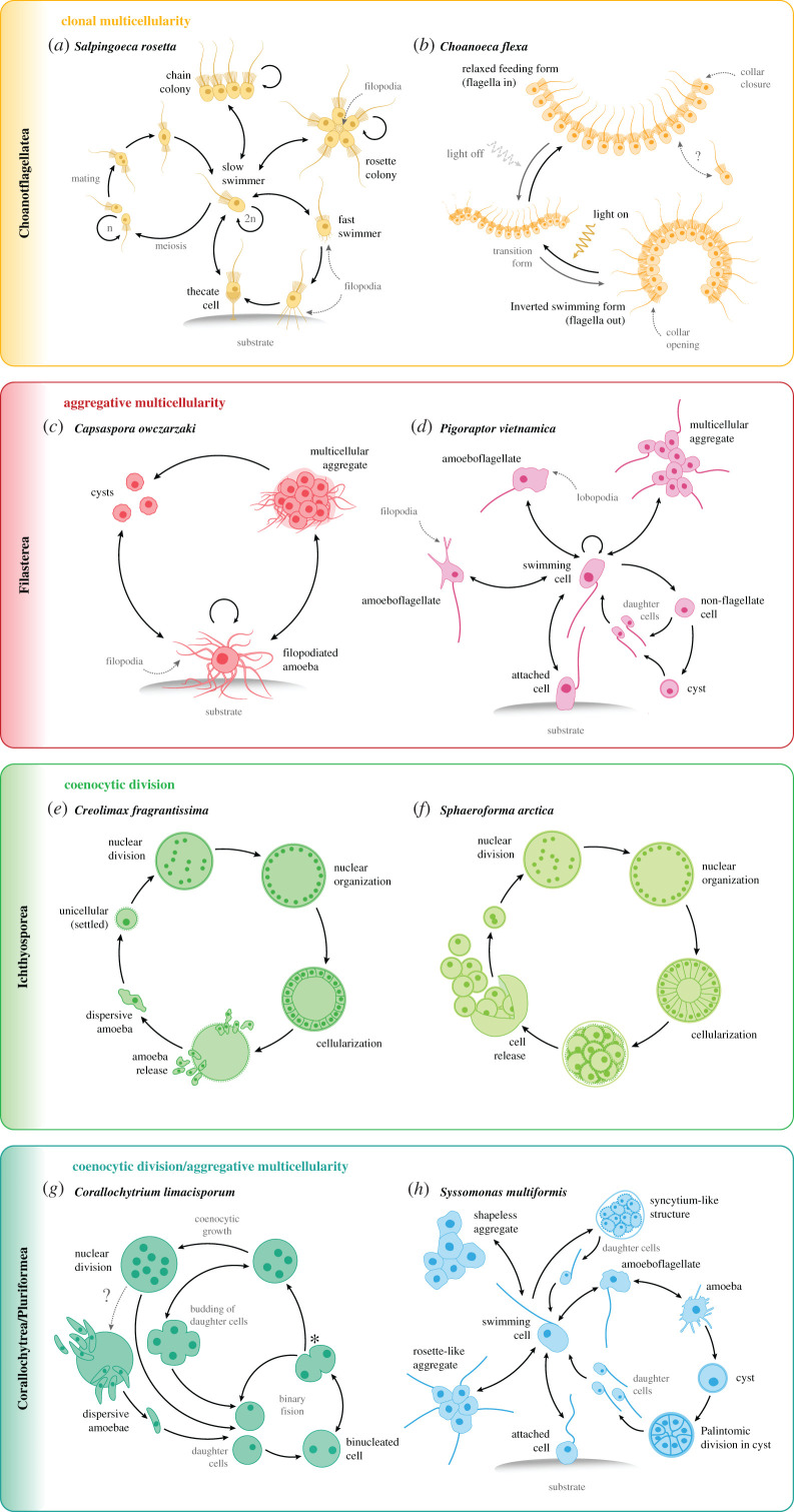
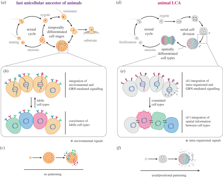
How animals evolved from a single-celled ancestor, transitioning from a unicellular lifestyle to a coordinated multicellular entity, remains a fascinating question. Key events in this transition involved the emergence of processes related to cell adhesion, cell-cell communication and gene regulation. To understand how these capacities evolved, we need to reconstruct the features of both the last common multicellular ancestor of animals and the last unicellular ancestor of animals. In this review, we summarize recent advances in the characterization of these ancestors, inferred by comparative genomic analyses between the earliest branching animals and those radiating later, and between animals and their closest unicellular relatives. We also provide an updated hypothesis regarding the transition to animal multicellularity, which was likely gradual and involved the use of gene regulatory mechanisms in the emergence of early developmental and morphogenetic plans. Finally, we discuss some new avenues of research that will complement these studies in the coming years.

Keywords: Holozoa; animal origins; cell-type evolution; evolutionary transitions; multicellularity.


Figures

Figure 1.
Figure 1.
Phylogenetic classification of animals and their unicellular relatives. (a) A timeline of different events during early animal evolution. The transition to animal multicellularity, and therefore the origin of the first animals, occurred sometime at the end of the Tonian period, according to molecular clock estimates. The oldest fossil or geological evidence of recognizable animals dates back to the Ediacaran period, with molecular clocks extending the emergence of different animal phyla back to the Cryogenian [–17]. Time units are million years ago (Ma). (b) Cladogram representing the major clades of the tree of animals and the major groups of unicellular relatives of animals: choanoflagellates, filastereans, ichthyosporeans and corallochytreans/pluriformeans. Coloured nodes indicate different ancestors that we can reconstruct and that are important to understand the transition to animal multicellularity; the highlighted internal branch (from the Urchoanozoan to the animal LCA) indicates the animal stem (see box 1; LCA = last common ancestor). Uncertain positions within the animal tree [–23] and within Holozoa [–26] are represented with polytomies.
Figure 2.
Figure 2.
An inferred gene repertoire of the last unicellular ancestor and the last common ancestor of animals. (a) The reconstruction of the last unicellular ancestor of animals is based on the presence of key metazoan genes in the genomes of unicellular relatives of animals. (b) Inferred gains present in the last common ancestor (LCA) of animals. Yellow indicates genes that originated prior to the emergence of the Holozoa LCA (pre-holozoan origins); green, genes that originated in Holozoa prior to the animal LCA (Holozoa origins); red, animal-specific genes that originated at the root of animals (animal origins). bHLH, basic helix–loop–helix transcription factors; BRA, Brachyury; CSK, C-terminal Src kinase; DRFs, diaphanous-related formins; EPS8, epidermal growth factor receptor kinase substrate 8; ERM, Ezrin–Radixin–Moesin proteins; GPCRs, G protein-coupled receptors; GSK3, glycogen synthase kinase 3; HD, homeodomain; MAGUKs, membrane-associated guanylate kinases; MAPKs, mitogen-activated protein kinases; MEF2, myocyte-specific enhancer factor 2; NF-κB, nuclear factor-κB; PI3 K, phosphatidylinositol 3-kinase; RFX, regulatory factor X transcription factors; RTKs, receptor tyrosine kinases; STAT, signal transducer and activator of transcription; TALEs, three amino acid loop extensions; TFs, transcription factors; TGFß, transforming growth factor beta. Data from [,,,,,,–121].
Figure 3.
Figure 3.
Temporally alternating life cycles of unicellular holozoans. Each panel shows life stage transitions of two unicellular holozoan species representing each clade. Arrows indicate directionality of the transition. Loop arrows indicate cell division. Dotted arrows with question marks between stages indicate potential (unconfirmed) life-stage transitions. (a) Life stages of the colonial choanoflagellate Salpingoeca rosetta [176,187]. The asexual life cycle (on the right) includes a single-celled sessile thecate stage (adhered to the substrate), slow and fast swimming single-celled stages, and two types of clonal colonial stages (chain and rosette colonies), in which neighbouring cells are linked by intercellular bridges [–190]. Starvation triggers the S. rosetta sexual cycle (on the left), in which diploid cells (slow swimmers) undergo meiosis and recombination, and the resulting haploid cells (which can also divide asexually) mate anisogamously [176,178]. (b) Life stages of the colonial choanoflagellate Choanoeca flexa [96]. Light-to-dark transitions induce C. flexa colonies to rapidly and reversibly invert their curvature while maintaining contacts among neighbouring cells between their collar microvilli, alternating between two colony conformations. In response to light, colonies exhibit a relaxed (flagella-in) feeding form. In the absence of light, colonies transition to an inverted (flagella-out) swimming form. (c) Life stages of the filasterean Capsaspora owczarzaki [64,65,98]. In the trophic proliferative (filopodial) stage, cells are amoebae adhered to the substrate, extending several long, thin actin-based filopodia. These amoebas can detach from the substrate and actively aggregate in the aggregative or ‘multicellular’ stage, producing an extracellular matrix that presumably binds them together. In response to crowding or stress, cells from both the amoeba and the aggregative stages can encyst by retracting the filopodia into a cystic or resistance stage. (d) Putative life stages of the filasterean Pigoraptor vietnamica [26,70]. Swimming flagellated cells can form long, thin, sometimes branching filopodia that can attach to the substrate. Flagellated cells can sometimes present wide lobopodia. Flagellated cells can retract the flagellum and become roundish, to either divide into two daughter flagellated cells or transition to a cystic stage. This can, in turn, produce two flagellated daughter cells. Cells can also form easily disintegrating aggregations of cells and feed jointly. The life stages of Pigoraptor chileana are very similar to the ones of P. vietnamica, but P. chileana shows a much reduced capability to produce filopodia and lobopodia (both stages are extremely rare in P. chileana). (e) Life stages of the ichthyosporean Creolimax fragrantissima [45,77]. Single-nucleated amoebae disperse until they settle and encyst. The rounded cell undergoes multiple rounds of synchronous nuclear division (coenocytic division) without cytoplasmic division. Nuclei are later arranged at the periphery of the cell as a large central vacuole grows. Finally, the coenocyte cellularizes and new amoebas are released to start the cycle over again. (f) Life stages of the ichthyosporean Sphaeroforma arctica [99,180]. Single-nucleated cells undergo multiple rounds of synchronous nuclear division (coenocytic division) without cytoplasmic division. Nuclei are later arranged at the periphery of the cell. Finally, the coenocyte cellularizes, releasing a number of daughter cells to start the cycle over again. (g) Life stages of the corallochytrean Corallochytrium limacisporum [22,83,191]. Reproduction in C. limacisporum occurs mainly through binary fission (99% of the cases), during which a binucleated cell divides into two, symmetrical, uninucleate cells. Binucleate cells can form two lobes that can lead to cellular division (forming two monoucleate cells), or can reverse towards spherical cells. At this point (*), cells can transition to coenocytic growth (1% of the cases) and continue dividing their nuclei further forming quadrinucleated cells. Quadrinucleated cells can often form a clover-like shape (similar to bilobed cell), that generates either four mononucleate cells or returns to spherical shape and further divides to an eight, 12 and up to 32 nuclei coenocyte. Coenocytes can release dispersive amoebas to start the cycle over again. (h) Putative life stages of the pluriformean Syssomonas multiformis [26,70]. A swimming flagellated cell can temporarily attach to the substrate through the anterior part of the cell body or move to the bottom and transform to an amoeboflagellate form by producing both wide lobopodia and thin short filopodia. Flagellated cells can lose the flagellum via different modes and transition into an amoeba stage, which produces thin, relatively short filopodia. Both amoeboflagellate and amoeba stages can transition back to the flagellate stage. Amoeboid cells can also encyst by retracting their filopodia and rounding the cell body. Palintomic divisions may occur in the cystic stage to release several flagellated daughter cells. Flagellated cells can partially merge and form temporary shapeless cell aggregates of both flagellated or non-flagellated cells and rosette-like colonies composed by only flagellated cells (showing outwards-directed flagella). In rich medium, solitary flagellated cells can sometimes actively merge and form a syncytium-like structure, which undergoes budding and releases flagellated daughter cells.
Figure 4.
Figure 4.
Our current perspective on important changes in the origin of animals. (a) The last unicellular ancestor of animals likely possessed a life cycle comprising different temporally regulated stages, including a sexually reproductive stage and at least one multicellular stage. (b) Cells within this multicellular structure were able to respond to different environmental stimuli thanks to a complex repertoire of signalling molecules and gene regulatory networks (GRNs), transitioning to labile cell stages. (c) This multicellular entity might have had a certain ability to integrate positional information from within the structure but lacked any axial/positional patterning. (d) The transition to animal origins likely involved some changes in this life cycle, already present by the time of the last common ancestor (LCA) of animals. (e) Cells within the multicellular structure acquired the ability to integrate spatial information from within the organism by making use of morphogenetic tools (such as ligands, receptors, and GRNs) (d′), which allowed the spatial organization of cell types (d″). Concomitantly, this developmental programme was conjoined with the sexual reproduction programme, by which gamete fusion was able to trigger the formation of a multicellular structure through serial division. (f) A greater ability to establish different cell types independently of the environment translates into the emergence of rudimentary morphogenetic plans, consisting of simple positional patterns (such as a primary axis) where different cell types localize to different regions of the organism (axial/positional patterning). It is worth emphasizing that the visual depictions presented here are mere representations of general concepts, and that we are by no means taking positions regarding specific details, such as the real structure of the life cycles, the number of cells, genes, molecules and GRNs implicated, the axial patterning or the morphological details of these organisms.

References

    1. Adl SM, et al. 2019. Revisions to the classification, nomenclature, and diversity of eukaryotes. J. Eukaryot. Microbiol. 66, 4-119. (10.1111/jeu.12691) - DOI - PMC - PubMed
    1. Baran Y, et al. 2019. MetaCell: analysis of single-cell RNA-seq data using K-nn graph partitions. Genome Biol. 20, 206. (10.1186/s13059-019-1812-2) - DOI - PMC - PubMed
    1. Burton PM. 2008. Insights from diploblasts; the evolution of mesoderm and muscle. J. Exp. Zoolog. B Mol. Dev. Evol. 310B, 5-14. (10.1002/jez.b.21150) - DOI - PubMed
    1. Chang C, Hemmati-Brivanlou A. 1998. Cell fate determination in embryonic ectoderm. J. Neurobiol. 36, 128-151. (10.1002/(SICI)1097-4695(199808)36:2<128::AID-NEU3>3.0.CO;2-3) - DOI - PubMed
    1. Lewis SL, Tam PPL. 2006. Definitive endoderm of the mouse embryo: formation, cell fates, and morphogenetic function. Dev. Dyn. 235, 2315-2329. (10.1002/dvdy.20846) - DOI - PubMed

Publication types