. 2021 Feb;11(2):200339.
doi: 10.1098/rsob.200339. Epub 2021 Feb 24.

An early Sox2-dependent gene expression programme required for hippocampal dentate gyrus development

Affiliations

An early Sox2-dependent gene expression programme required for hippocampal dentate gyrus development

Sara Mercurio et al. Open Biol. 2021 Feb.

Abstract

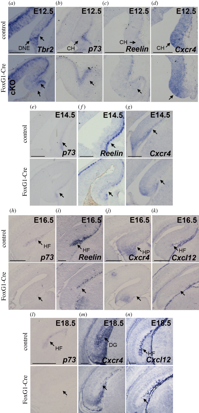
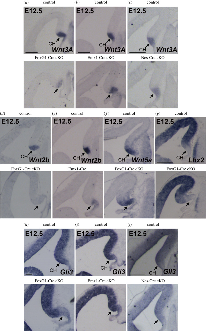
The hippocampus is a brain area central for cognition. Mutations in the human SOX2 transcription factor cause neurodevelopmental defects, leading to intellectual disability and seizures, together with hippocampal dysplasia. We generated an allelic series of Sox2 conditional mutations in mouse, deleting Sox2 at different developmental stages. Late Sox2 deletion (from E11.5, via Nestin-Cre) affects only postnatal hippocampal development; earlier deletion (from E10.5, Emx1-Cre) significantly reduces the dentate gyrus (DG), and the earliest deletion (from E9.5, FoxG1-Cre) causes drastic abnormalities, with almost complete absence of the DG. We identify a set of functionally interconnected genes (Gli3, Wnt3a, Cxcr4, p73 and Tbr2), known to play essential roles in hippocampal embryogenesis, which are downregulated in early Sox2 mutants, and (Gli3 and Cxcr4) directly controlled by SOX2; their downregulation provides plausible molecular mechanisms contributing to the defect. Electrophysiological studies of the Emx1-Cre mouse model reveal altered excitatory transmission in CA1 and CA3 regions.

Keywords: Sox; Sox2; gene regulation; mouse genetic models; transcription factors.


Figures

Figure 1.
Figure 1.
Sox2 expression in the dorsal telencephalon. (a) Schematic of the development of the hippocampus in the dorsal telencephalon. (be) ISH for Sox2 on coronal section of mouse brains at E10.5 (b) E12.5 (c), E15.5 (d) and E18.5 (e). Arrows indicate Sox2 expression in the developing hippocampus in particular in the dorsal telencephalon in (b), in the CH in (c), in the dorsal migratory stream (DMS) in (d) and in the DG in (e′). (fi) IF of Sox2 (fi), of markers of CRC, Reelin (f,g) and P73 (i), and of a marker of differentiating neurons TuJ1 (h). Representative single optical confocal sections are shown. Scale bars 200 µm. CH, cortical hem; DNE, dentate neuroepithelium; HNE, hippocampal neuroepithelium; DMS, dentate migratory stream; HF, hippocampal fissure; DG, dentate gyrus; F, fimbria; Th, thalamus.
Figure 2.
Figure 2.
Hippocampal DG development is impaired in FoxG1-Cre cKO Sox2 mutants. (a) GFAP IF at E18.5 on coronal sections of control and FoxG1-Cre cKO hippocampi (controls n = 7 (Sox2 +/+ n = 4, Sox2 +/−; FoxG1 +/− n = 3); mutants n = 4). (bd) ISH at E18.5 for NeuroD (b) (controls n = 4 (Sox2 +/+ n = 2, Sox2 +/−; FoxG1 +/− n = 2); mutants n = 3), Hes5 (c) (controls n = 2 (Sox2 +/−; FoxG1 +/− n = 2); mutants n = 2) and Prox1 (d) (controls n = 2; mutants n = 2) on coronal sections of control and FoxG1-Cre cKO hippocampi. Arrows indicate the underdeveloped DG in cKO. Scale bars, 200 µm.
Figure 3.
Figure 3.
Hippocampus development is affected by Sox2 loss, the earlier Sox2 is ablated the stronger the phenotype observed. ISH for Cadherin8 (ac), Reelin (df) and Tbr2 (gi) on coronal sections of control and Sox2 FoxG1-Cre cKO brains at E18.5 (a,d,g), control and Emx1-Cre cKO brains at P0 (b,e,h) and control and Nes-Cre cKO brains at P0 (c,f,i). At least three controls and three mutants were analysed for each probe. A schematic of the timing of Sox2 ablation with the different Cre lines is at the bottom. Scale bars, 200 µm. DG, dentate gyrus; HF, hippocampal fissure; DMS, dentate migratory stream; Tel, telencephalon; dTel, dorsal telencephalon; vTel, ventral telencephalon; CH, cortical hem.
Figure 4.
Figure 4.
Expression of genes important for the development of the hippocampus is affected by Sox2 loss in FoxG1-Cre cKO. (ad) ISH at E12.5 on coronal sections of control and FoxG1-Cre cKO dorsal telencephalons for Tbr2 (controls n = 10, mutants n = 10) (a), P73 (controls n = 3, mutants n = 3) (b), Reelin (controls n = 7, mutants n = 6) (c) and Cxcr4 (controls n = 7, mutants n = 7) (d). (eg) ISH at E14.5 on coronal sections of control and FoxG1-Cre cKO brains for P73 (controls n = 3, mutants n = 3) (e), Reelin (controls n = 7, mutants n = 5) (f) and Cxcr4 (controls n = 3, mutants n = 3) (g). (hk) ISH at E16.5 on coronal sections of control and FoxG1-Cre cKO hippocampi for P73 (controls n = 2, mutants n = 2) (h), Reelin (controls n = 6, mutants n = 5) (i), Cxcr4 (controls n = 5, mutants n = 4) (j) and Cxcl12 (controls n = 4, mutants n = 3) (k). (ln)) ISH at E18.5 on coronal sections of control and FoxG1-Cre cKO hippocampi for P73 (controls n = 3, mutants n = 3) (l), Cxcr4 (controls n = 5, mutants n = 4) (m) and Cxcl12 (controls n = 5, mutants n = 4) (n). Arrows indicate the downregulation of expression in the mutant CH, dentate neuroepithelium (DNE), hippocampal primordium (HP), DG and hippocampal fissure (HF). Scale bars, 200 µm.
Figure 5.
Figure 5.
Expression of key molecules for hippocampal development is downregulated in the CH of FoxG1-Cre cKO but mildly or not affected in Emx1-Cre or Nes-Cre cKO. ISH at E12.5 for Wnt3A (ac), Wnt2b (d,e), Wnt5A (f), Lhx2 (g) and Gli3 (hj) on control and FoxG1-Cre cKO (a,d,f,g,h), Emx1-Cre cKO (b,e,i) and Nes-Cre cKO (c,j) coronal brain sections. Arrows indicate the CH. At least three controls and three mutants were analysed for each probe. Scale bars, 200 µm.
Figure 6.
Figure 6.
SOX2 acts on distal enhancers and on long-range enhancer–promoter interactions of several genes key to hippocampal development, and activates a Gli3 intronic enhancer in a dose-dependent way. (a) Diagram of the Gli3 gene, and SOX2-binding profile across the Gli3 locus in NSC (ChIPseq data from [39]). A Sox2-dependent 80 kb long-range interaction connects the Gli3 promoter with a SOX2-bound region, in the second intron (ChIA-PET data from [39]). This region acts as a brain-specific enhancer in E10.5 mouse embryo (image from https://enhancer.lbl.gov/); it was cloned into the depicted luciferase vector, upstream to a minimal tk promoter, to address its responsivity to Sox2. (b) Enhancer activation assay in Neuro2a cells transfected with the constructs in (a): Gli3 enhancer + tk promoter (blue histograms), or tk promoter only (grey histograms). Cotransfection of these constructs with increasing amounts of a Sox2-expressing vector (Sox2, X-axis), but not of a control ‘empty’ vector (empty Sox2), or a Mash1-expressing vector (Mash1), resulted in dose-dependent increase of luciferase activity (Y-axis) driven by the Gli3 enhancer + tk-prom vector, but not the tk-prom only vector. The molar ratios, compared with the luciferase vector (set at 1) were: +, 1 : 0.050; ++, 1 : 0.075; +++, 1 : 0.125; ++++, 1 : 0.25; +++++, 1 : 0.5. Results are represented as fold-change increase in activity compared with the tk-prom only vector, which is set at 1. Values are the mean of two (for Sox2+ and Sox2++) or three (other samples) independent experiments carried out in triplicate. Error bars represent s.d. (c) Diagram of the Cxcr4 gene, reporting SOX2 binding and Sox2-dependent long-range interactions in NSC (as in (a) for Gli3; data from [39]). Note that the Cxcr4 promoter is connected to a SOX2-bound region within the intron of a different gene, Dars; this region acts as a brain-specific enhancer in transgenic zebrafish embryos (picture from [39]). (d) A model depicting the activation, by Sox2, of different genes key to hippocampal development (present paper), some of which cross-regulate each other; in red, direct SOX2 targets; in bold, early expressed hippocampal regulators, downregulated already at early stages in Sox2 mutants (see Discussion).
Figure 7.
Figure 7.
In Emx1-Cre cKO mice, excitatory transmission is altered in CA3 and CA1 hippocampal regions. Early Sox2 ablation leads to alterations in the excitatory input onto both CA3 and CA1 pyramidal neurons. (a) Typical firing response to a 200 pA stimulus of injected current in a CA3 pyramidal neuron. (b) Average stimulus/frequency relation for hippocampal pyramidal neurons recorded in CA3 (circles) and CA1 (squares). No major differences were observed between control (black) and mutant animals (red). (c,d) EPSCs traces, at −68 mV, recorded in simulated physiologic conditions onto pyramidal neurons in CA3 and CA1 region, respectively. Insets. Magnification of a representative EPSC event. (e) Average EPSCs frequencies and median amplitudes observed in CA3 pyramidal neurons recorded from 15 animals between p19 and p31. In mutant animals, Sox2 ablation induced a significant increase in EPSCs frequency compared to controls (8.60 ± 1.15 Hz, n = 20 and 5.59 ± 0.57 Hz, n = 28, respectively; p = 0.03941, with Mann–Whitney test), whereas no significant effect was produced on event amplitudes (9.21 ± 0.47 pA, n = 20 and 10.01 ± 0.61 pA, n = 28, respectively). (f) Amplitude distribution of the total amount of collected EPSCs showing no major differences between control and mutant mice. (g) In CA1, EPSCs frequency significantly decreased in mutant animals compared to controls (1.46 ± 0.35 Hz, n = 13 and 2.91 ± 0.52 Hz, n = 13, respectively; p = 0.02745, with Mann–Whitney test). No difference in the average median amplitude was observed (controls: 7.75 ± 0,69 pA, n = 20 and mutants: 6.92 ± 0.44 pA, n = 28). (h) The amplitude distribution of the total pool of events recorded from 13 animals between p19 and p31 showed no major alterations between control and mutant mice.

Similar articles

Cited by

References

    1. Kandel ER, Schwartz JH, Jessell TM. 2000. Principles of neural science, 4th edn. New York, NY: McGraw-Hill, Health Professions Division.
    1. Berg DA, et al. 2019. A common embryonic origin of stem cells drives developmental and adult neurogenesis. Cell 177, 654-668 e15. (10.1016/j.cell.2019.02.010) - DOI - PMC - PubMed
    1. Zhong S, et al. 2020. Decoding the development of the human hippocampus. Nature 577, 531-536. (10.1038/s41586-019-1917-5) - DOI - PubMed
    1. Fantes J, et al. 2003. Mutations in SOX2 cause anophthalmia. Nat. Genet. 33, 461-463. (10.1038/ng1120) - DOI - PubMed
    1. Kondoh H L-BReb. 2016. Sox2, biology and role in development and disease. London, UK: Academic Press.

Publication types

MeSH terms

LinkOut - more resources