

MRI-based radiomics approach for differentiation of hypovascular non-functional pancreatic neuroendocrine tumors and solid pseudopapillary neoplasms of the pancreas
- PMID: 33622277
- PMCID: PMC7901077
- DOI: 10.1186/s12880-021-00563-x
MRI-based radiomics approach for differentiation of hypovascular non-functional pancreatic neuroendocrine tumors and solid pseudopapillary neoplasms of the pancreas
Abstract
Background: This study aims to investigate the value of radiomics parameters derived from contrast enhanced (CE) MRI in differentiation of hypovascular non-functional pancreatic neuroendocrine tumors (hypo-NF-pNETs) and solid pseudopapillary neoplasms of the pancreas (SPNs).
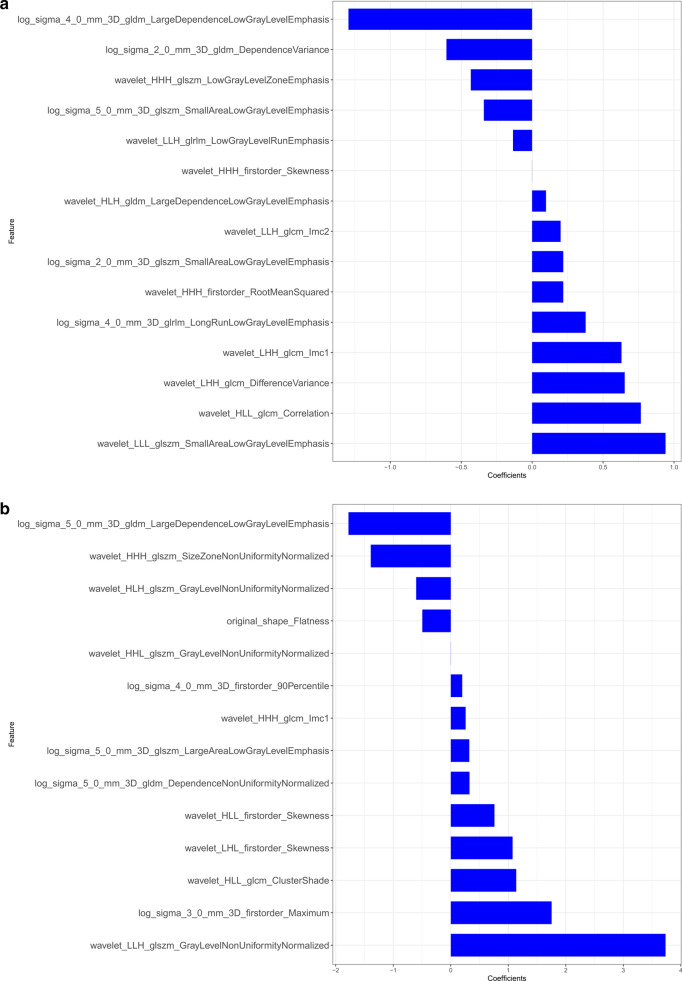
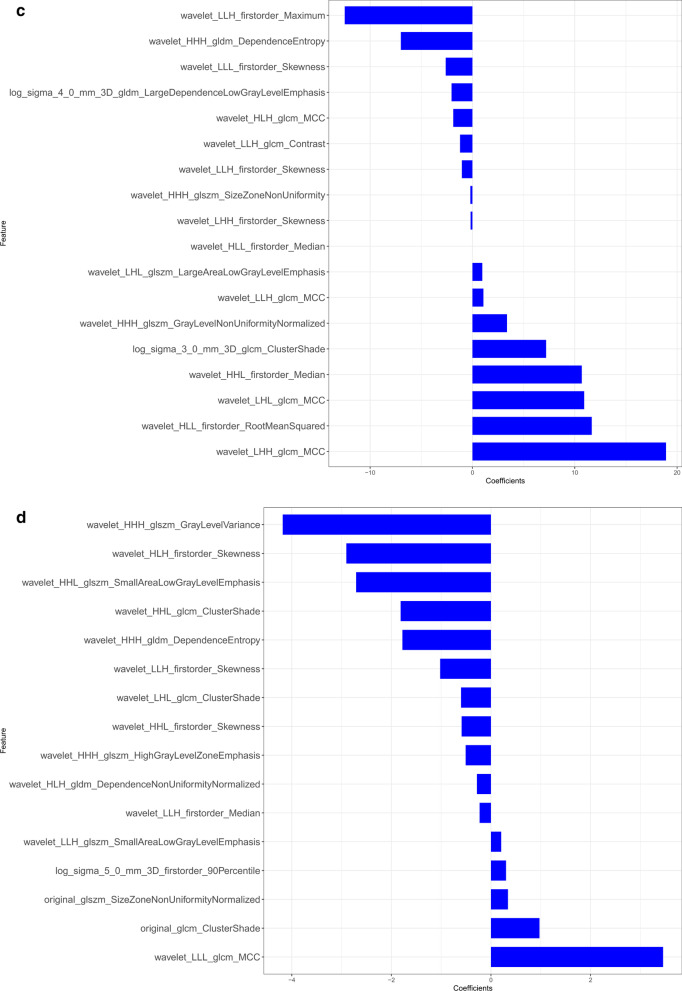
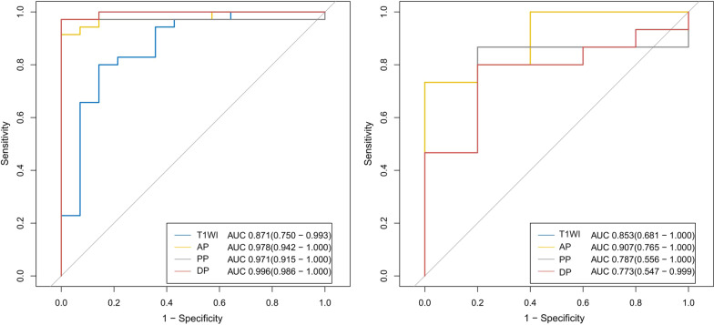
Methods: Fifty-seven SPN patients and twenty-two hypo-NF-pNET patients were enrolled. Radiomics features were extracted from T1WI, arterial, portal and delayed phase of MR images. The enrolled patients were divided into training cohort and validation cohort with the 7:3 ratio. We built four radiomics signatures for the four phases respectively and ROC analysis were used to select the best phase to discriminate SPNs from hypo-NF-pNETs. The chosen radiomics signature and clinical independent risk factors were integrated to construct a clinic-radiomics nomogram.
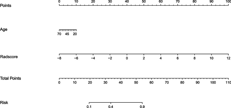
Results: SPNs occurred in younger age groups than hypo-NF-pNETs (P < 0.0001) and showed a clear preponderance in females (P = 0.0185). Age was a significant independent factor for the differentiation of SPNs and hypo-NF-pNETs revealed by logistic regression analysis. With AUC values above 0.900 in both training and validation cohort (0.978 [95% CI, 0.942-1.000] in the training set, 0.907 [95% CI, 0.765-1.000] in the validation set), the radiomics signature of the arterial phase was picked to build a clinic-radiomics nomogram. The nomogram, composed by age and radiomics signature of the arterial phase, showed sufficient performance for discriminating SPNs and hypo-NF-pNETs with AUC values of 0.965 (95% CI, 0.923-1.000) and 0.920 (95% CI, 0.796-1.000) in the training and validation cohorts, respectively. Delong Test did not demonstrate statistical significance between the AUC of the clinic-radiomics nomogram and radiomics signature of arterial phase.
Conclusion: CE-MRI-based radiomics approach demonstrated great potential in the differentiation of hypo-NF-pNETs and SPNs.
Keywords: Magnetic resonance imaging; Pancreatic neuroendocrine tumors; Radiomics; Solid pseudopapillary neoplasms of the pancreas.
Conflict of interest statement
The authors declare that they have no competing interests.
Figures


Similar articles
-
How does the pancreatic solid pseudopapillary neoplasm confuse us: Analyzing from the point view of MRI-based radiomics?Magn Reson Imaging. 2022 Jan;85:38-43. doi: 10.1016/j.mri.2021.10.034. Epub 2021 Oct 20. Magn Reson Imaging. 2022. PMID: 34687847
-
Radiomics analysis from magnetic resonance imaging in predicting the grade of nonfunctioning pancreatic neuroendocrine tumors: a multicenter study.Eur Radiol. 2024 Jan;34(1):90-102. doi: 10.1007/s00330-023-09957-7. Eur Radiol. 2024. PMID: 37552258 Free PMC article.
-
CT radiomics may predict the grade of pancreatic neuroendocrine tumors: a multicenter study.Eur Radiol. 2019 Dec;29(12):6880-6890. doi: 10.1007/s00330-019-06176-x. Epub 2019 Jun 21. Eur Radiol. 2019. PMID: 31227882
-
Radiomics in CT and MR imaging of the liver and pancreas: tools with potential for clinical application.Abdom Radiol (NY). 2024 Jan;49(1):322-340. doi: 10.1007/s00261-023-04071-0. Epub 2023 Oct 27. Abdom Radiol (NY). 2024. PMID: 37889265 Review.
-
The Role of MRI in the Diagnosis of Solid Pseudopapillary Neoplasm of the Pancreas and Its Mimickers: A Case-Based Review with Emphasis on Differential Diagnosis.Diagnostics (Basel). 2023 Mar 13;13(6):1074. doi: 10.3390/diagnostics13061074. Diagnostics (Basel). 2023. PMID: 36980388 Free PMC article. Review.
Cited by
-
Using Quantitative Imaging for Personalized Medicine in Pancreatic Cancer: A Review of Radiomics and Deep Learning Applications.Cancers (Basel). 2022 Mar 24;14(7):1654. doi: 10.3390/cancers14071654. Cancers (Basel). 2022. PMID: 35406426 Free PMC article. Review.
-
Can solid pseudopapillary neoplasm of the pancreas without degeneration be diagnosed with imaging? a comparison study of the solid component of solid pseudopapillary neoplasm, neuroendocrine neoplasm, and ductal adenocarcinoma.Abdom Radiol (NY). 2023 Mar;48(3):936-951. doi: 10.1007/s00261-023-03814-3. Epub 2023 Jan 28. Abdom Radiol (NY). 2023. PMID: 36708377
-
Development and validation of an ultrasound-based prediction model for differentiating between malignant and benign solid pancreatic lesions.Eur Radiol. 2022 Dec;32(12):8296-8305. doi: 10.1007/s00330-022-08930-0. Epub 2022 Jun 25. Eur Radiol. 2022. PMID: 35751698 Free PMC article.
-
Comparison between solid pseudopapillary neoplasms of the pancreas and pancreatic ductal adenocarcinoma with cystic changes using computed tomography.World J Radiol. 2024 Jun 28;16(6):211-220. doi: 10.4329/wjr.v16.i6.211. World J Radiol. 2024. PMID: 38983836 Free PMC article.
-
Kidney Cancer Prediction Empowered with Blockchain Security Using Transfer Learning.Sensors (Basel). 2022 Oct 2;22(19):7483. doi: 10.3390/s22197483. Sensors (Basel). 2022. PMID: 36236584 Free PMC article.
References
Publication types
MeSH terms
Supplementary concepts
LinkOut - more resources
Full Text Sources
Other Literature Sources
Medical
