

Dermatofibrosarcoma protuberans in a young patient with epidermolysis bullosa: a case report
- PMID: 33622311
- PMCID: PMC7903690
- DOI: 10.1186/s12893-021-01105-6
Dermatofibrosarcoma protuberans in a young patient with epidermolysis bullosa: a case report
Abstract
Background: Epidermolysis bullosa is a group of rare inherited skin diseases characterized by blister formation following mechanical skin trauma. Epidermolysis bullosa is associated with increased skin cancer rates, predominantly squamous cell carcinomas, yet to our best knowledge, there is no reported case of dermatofibrosarcoma protuberans in a patient with Epidermolysis bullosa.
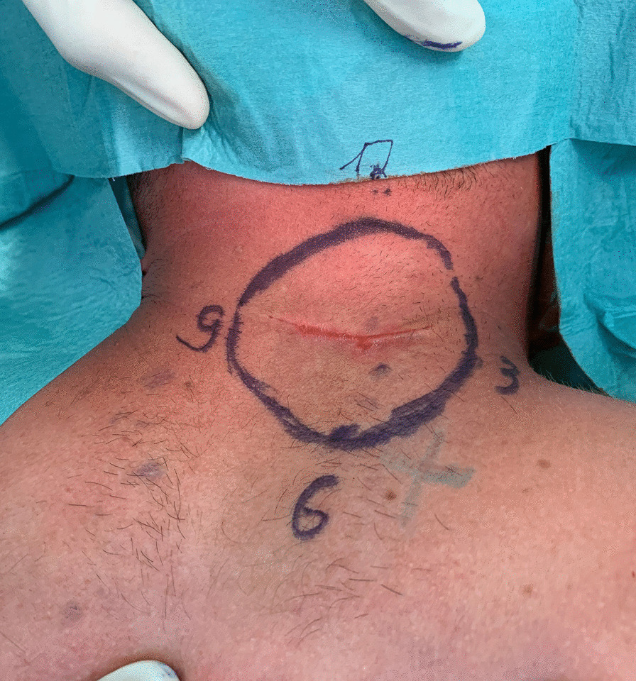
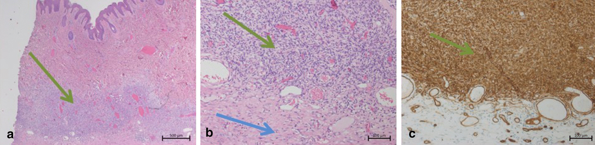
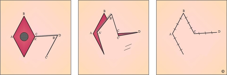
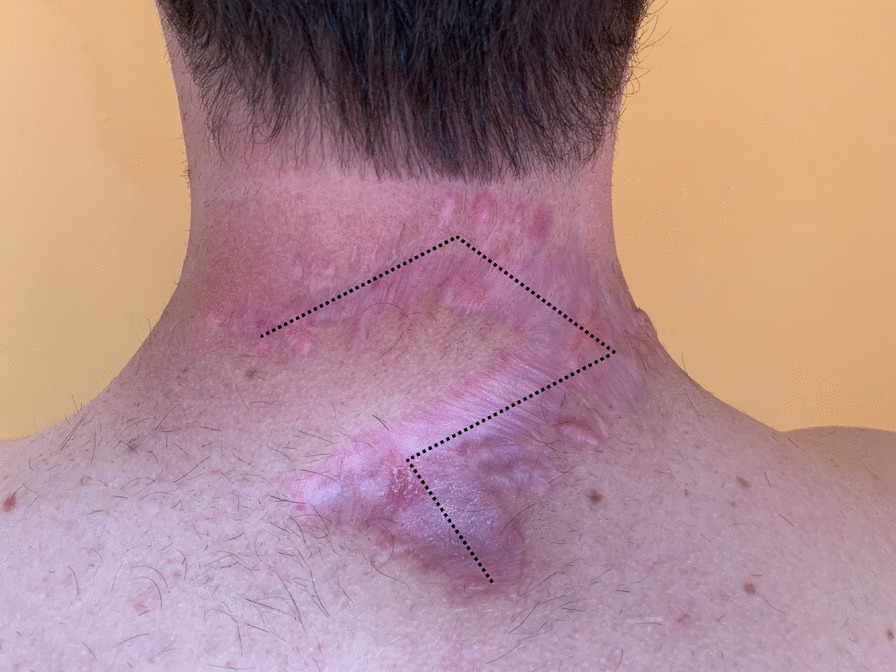
Case presentation: Here, we present a 26-year-old man with junctional epidermolysis bullosa, who developed a DFSP on the neck. Initial, the skin alteration was mistakenly not considered malignant, which resulted in inadequate safety margins. The complete resection required a local flap to close the defect, which is not unproblematic because of the chronic inflammation and impaired healing potential of the skin due to Epidermolysis bullosa.
Conclusions: To our best knowledge, this is the first reported case of a skin-associated sarcoma in a patient with EB; however, further investigation is required to verify a correlation.
Keywords: Case report; Dermatofibrosarcoma protuberans; Epidermolysis bullosa; Local skin flap; Reconstructive surgery.
Conflict of interest statement
The authors declare they have no competing interests.
Figures
Similar articles
-
Single-institution review of managing dermatofibrosarcoma protuberans.ANZ J Surg. 2016 May;86(5):372-6. doi: 10.1111/ans.13276. Epub 2015 Sep 2. ANZ J Surg. 2016. PMID: 26334110
-
Generalized atrophic benign epidermolysis bullosa in 2 siblings complicated by multiple squamous cell carcinomas.Arch Dermatol. 1998 Feb;134(2):199-203. doi: 10.1001/archderm.134.2.199. Arch Dermatol. 1998. PMID: 9487212
-
A Rare Case of Misdiagnosis: Recurrence of Dermatofibrosarcoma Protuberans That Was Treated Surgicallyas a Keloid.Med Arch. 2018 Feb;72(1):74-75. doi: 10.5455/medarh.2018.72.74-75. Med Arch. 2018. PMID: 29416224 Free PMC article.
-
Dermatofibrosarcoma protuberans: a rare entity and review of the literature.J BUON. 2014 Jan-Mar;19(1):34-41. J BUON. 2014. PMID: 24659640 Review.
-
Dermatofibrosarcoma protuberans in children and adolescents: Clinical presentation, histology, treatment, and review of the literature.J Plast Reconstr Aesthet Surg. 2014 Sep;67(9):1222-9. doi: 10.1016/j.bjps.2014.05.031. Epub 2014 Jun 2. J Plast Reconstr Aesthet Surg. 2014. PMID: 24973861 Review.
References
-
- Bruckner-Tuderman L. Epidermolysis bullosa: eine interdisziplinäre Herausforderung. Dtsch Ärztebl. 2001;8:5.
Publication types
MeSH terms
LinkOut - more resources
Full Text Sources
Other Literature Sources
Medical
