. 2021 Feb 23;8(1):16.
doi: 10.1186/s40779-021-00309-4.

Toll-like receptor 5-mediated signaling enhances liver regeneration in mice

Affiliations

Toll-like receptor 5-mediated signaling enhances liver regeneration in mice

Wen Zhang et al. Mil Med Res. .

Abstract

Background: Toll-like receptor 5 (TLR5)-mediated pathways play critical roles in regulating the hepatic immune response and show hepatoprotective effects in mouse models of hepatic diseases. However, the role of TLR5 in experimental models of liver regeneration has not been reported. This study aimed to investigate the role of TLR5 in partial hepatectomy (PHx)-induced liver regeneration.

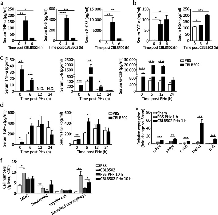
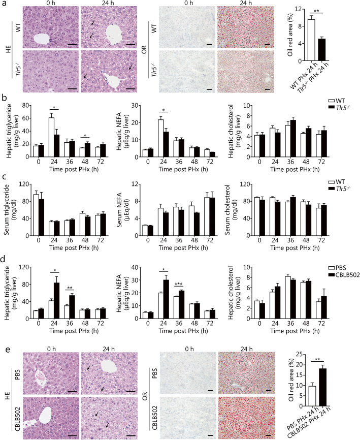
Methods: We performed 2/3 PHx in wild-type (WT) mice, TLR5 knockout mice, or TLR5 agonist CBLB502 treated mice, as a model of liver regeneration. Bacterial flagellin content was measured with ELISA, and hepatic TLR5 expression was determined with quantitative PCR analyses and flow cytometry. To study the effects of TLR5 on hepatocyte proliferation, we analyzed bromodeoxyuridine (BrdU) incorporation and proliferating cell nuclear antigen (PCNA) expression with immunohistochemistry (IHC) staining. The effects of TLR5 during the priming phase of liver regeneration were examined with quantitative PCR analyses of immediate early gene mRNA levels, and with Western blotting analysis of hepatic NF-κB and STAT3 activation. Cytokine and growth factor production after PHx were detected with real-time PCR and cytometric bead array (CBA) assays. Oil Red O staining and hepatic lipid concentrations were analyzed to examine the effect of TLR5 on hepatic lipid accumulation after PHx.

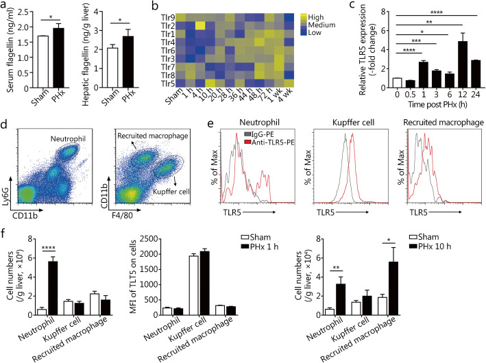
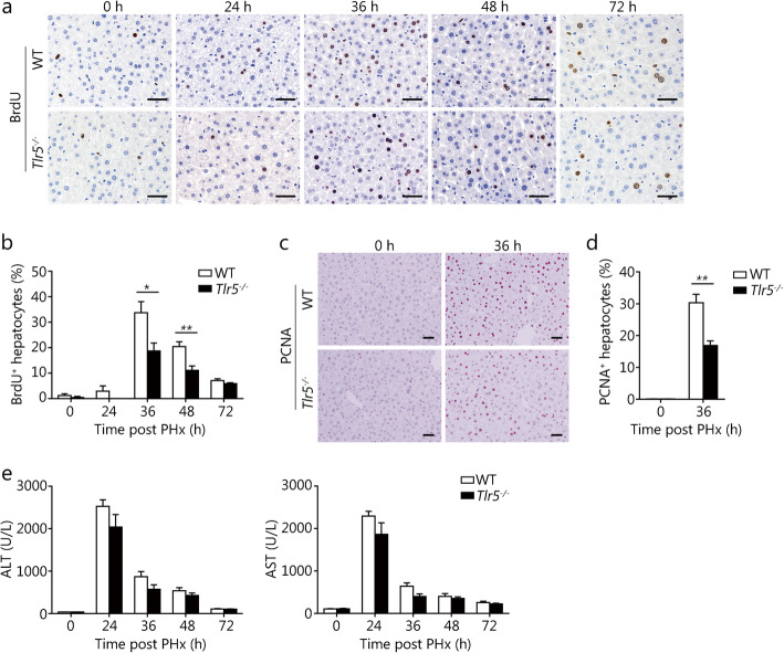
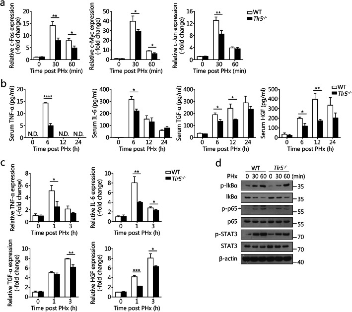
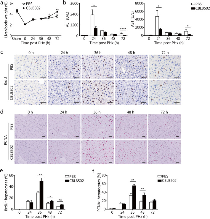
Results: The bacterial flagellin content in the serum and liver increased, and the hepatic TLR5 expression was significantly up-regulated in WT mice after PHx. TLR5-deficient mice exhibited diminished numbers of BrdU- and PCNA-positive cells, suppressed immediate early gene expression, and decreased cytokine and growth factor production. Moreover, PHx-induced hepatic NF-κB and STAT3 activation was inhibited in Tlr5-/- mice, as compared with WT mice. Consistently, the administration of CBLB502 significantly promoted PHx-mediated hepatocyte proliferation, which was correlated with enhanced production of proinflammatory cytokines and the recruitment of macrophages and neutrophils in the liver. Furthermore, Tlr5-/- mice displayed significantly lower hepatic lipid concentrations and smaller Oil Red O positive areas than those in control mice after PHx.

Conclusion: We reveal that TLR5 activation contributes to the initial events of liver regeneration after PHx. Our findings demonstrate that TLR5 signaling positively regulates liver regeneration and suggest the potential of TLR5 agonist to promote liver regeneration.

Keywords: CBLB502; Liver regeneration; NF-κB; Partial hepatectomy; Toll-like receptor 5.


Conflict of interest statement

The authors declare that they have no competing interests.

Figures

Fig. 1
Fig. 1
TLR5 is up-regulated in the liver after PHx. a Flagellin concentrations in the serum and liver in WT mice 6 h after sham surgery or PHx. b TLR expression levels in regenerating WT mouse liver after PHx from a published transcriptome dataset (GSE95135). c Relative mRNA levels of hepatic TLR5 at the indicated times after PHx. Flow cytometry gating strategy (d) and TLR5 expression levels (e) of liver neutrophils, KCs, and recruited macrophages in WT mice. f Cell counts and mean fluorescence intensity of TLR5 on liver neutrophils, KCs, and recruited macrophages at the indicated times after PHx. Panel a shows means ± SD. Data are representative of three independent experiments. Panels c and f show means ± SEM. Data are derived from two or three independent experiments with similar results. n = 3–6. *P < 0.05, **P < 0.01, ***P < 0.001, ****P < 0.0001
Fig. 2
Fig. 2
TLR5 deficiency attenuates hepatocyte proliferation after PHx. Proliferation was measured with BrdU and PCNA immunohistochemistry staining. Representative IHC staining images were shown (a and c), and the percentages of BrdU-positive cells (b) and PCNA-positive cells (d) were counted. e Serum ALT and AST levels in WT and Tlr5−/− mice subjected to PHx. Results are expressed as the mean ± SEM. Data are derived from two or three independent experiments with similar results. n = 5–6. *P < 0.05, **P < 0.01
Fig. 3
Fig. 3
TLR5 deficiency suppresses hepatocyte priming in PHx-induced liver regeneration. a Relative mRNA levels of hepatic c-fos, c-myc, and c-jun in WT and Tlr5−/− mice at the indicated times after PHx. b Serum TNF-α, IL-6, TGF-α, and HGF were measured in regenerating WT and Tlr5−/− mouse livers. c Quantification of hepatic TNF-α, IL-6, TGF-α, and HGF mRNA expression in WT and Tlr5−/− mice at the indicated times after PHx. d Western blot analysis of the indicated target proteins in WT and Tlr5−/− mouse livers after PHx, with β-actin as the loading control. Panels a and c show means ± SD. Results are representative of three independent experiments. Panel b shows means ± SEM. Data are derived from two or three independent experiments with similar results. n = 4–6. Western blot data in panel d are representative of three independent experiments. N.D., not detected. *P < 0.05, **P < 0.01, ***P < 0.001, ****P < 0.0001
Fig. 4
Fig. 4
The TLR5 agonist CBLB502 enhances hepatocyte proliferation in mice after PHx. Liver to body weight ratio (a), and serum ALT and AST levels (b) in PBS and CBLB502 treated mice after PHx. Hepatocyte proliferation was measured with BrdU (c) and PCNA (d) immunohistochemistry staining. Percentages of BrdU-positive cells (e) and PCNA-positive cells (f) were counted at the indicated times after PHx. Results are expressed as the mean ± SEM. Data are derived from two or three independent experiments with similar results. n = 6–12. *P < 0.05, **P < 0.01
Fig. 5
Fig. 5
Enhanced inflammatory response in CBLB502-pretreated mice after PHx. Serum TNF-α, IL-6, and G-CSF (a), and TGF-α and HGF (b) in CBLB502 treated mice at the indicated times. Serum TNF-α, IL-6, and G-CSF (c), and TGF-α and HGF (d) were measured in regenerating PBS and CBLB502 treated mouse livers after PHx. e Relative mRNA levels of hepatic c-Fos, c-Myc, c-Jun, TNF-α, and IL-6 in PBS and CBLB502 treated mice after PHx. f Cell counts of liver MNCs, neutrophils, KCs, and recruited macrophages in PBS and CBLB502 treated mice after sham surgery or PHx. Panel e shows means ± SD. Data are representative of three independent experiments. Panels a-d, f show means ± SEM. Data are derived from two or three independent experiments with similar results. n = 3–7. N.D., not detected. *P < 0.05, **P < 0.01, ***P < 0.001, ****P < 0.0001
Fig. 6
Fig. 6
TLR5 signaling contributes to hepatic lipid accumulation induced by PHx. a H&E staining, Oil Red O staining, and quantification of positive areas of WT and Tlr5−/− mouse livers after PHx. Triglyceride, NEFA, and cholesterol levels in liver extracts (b) and serum (c) were measured in WT and Tlr5−/− mice at the indicated times after PHx. d Analysis of hepatic triglyceride, NEFA, and cholesterol levels in PBS and CBLB502 treated mice after PHx. e H&E staining, Oil Red O staining, and quantification of positive areas of PBS and CBLB502 treated mouse livers after PHx. Black arrows indicate hepatic lipids. Results are expressed as the mean ± SEM. Data are derived from two or three independent experiments with similar results. n = 5–7. *P < 0.05, **P < 0.01, ***P < 0.001

References

    1. Preziosi ME, Monga SP. Update on the mechanisms of liver regeneration. Semin Liver Dis. 2017;37(2):141–151. doi: 10.1055/s-0037-1601351. - DOI - PMC - PubMed
    1. Tao Y, Wang M, Chen E, Tang H. Liver regeneration: analysis of the main relevant signaling molecules. Mediat Inflamm. 2017;2017:4256352. - PMC - PubMed
    1. Mitchell C, Gilgenkrantz H. Transcriptional profiling of liver regeneration: new approaches to an old trick! J Hepatol. 2003;38(6):847–849. doi: 10.1016/S0168-8278(03)00158-2. - DOI - PubMed
    1. Iimuro Y, Fujimoto J. TLRs, NF-kappaB, JNK, and liver regeneration. Gastroenterol Res Pract. 2010;2010:598109. doi: 10.1155/2010/598109. - DOI - PMC - PubMed
    1. Shi JH, Line PD. Hallmarks of postoperative liver regeneration: an updated insight on the regulatory mechanisms. J Gastroenterol Hepatol. 2020;35(6):960–966. doi: 10.1111/jgh.14944. - DOI - PubMed

Publication types

LinkOut - more resources