. 2021 Feb 23;9(1):30.
doi: 10.1186/s40478-021-01129-2.

Frontotemporal lobar degeneration proteinopathies have disparate microscopic patterns of white and grey matter pathology

Affiliations

Frontotemporal lobar degeneration proteinopathies have disparate microscopic patterns of white and grey matter pathology

Lucia A A Giannini et al. Acta Neuropathol Commun. .

Abstract

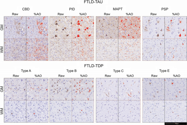
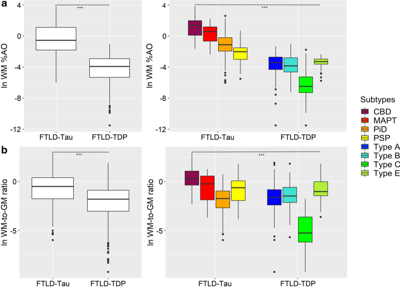
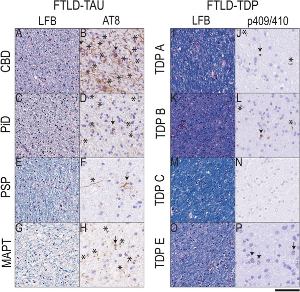
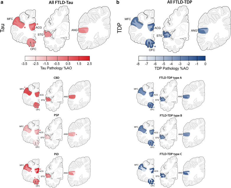
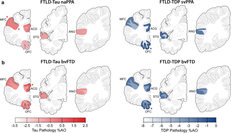
Frontotemporal lobar degeneration proteinopathies with tau inclusions (FTLD-Tau) or TDP-43 inclusions (FTLD-TDP) are associated with clinically similar phenotypes. However, these disparate proteinopathies likely differ in cellular severity and regional distribution of inclusions in white matter (WM) and adjacent grey matter (GM), which have been understudied. We performed a neuropathological study of subcortical WM and adjacent GM in a large autopsy cohort (n = 92; FTLD-Tau = 37, FTLD-TDP = 55) using a validated digital image approach. The antemortem clinical phenotype was behavioral-variant frontotemporal dementia (bvFTD) in 23 patients with FTLD-Tau and 42 with FTLD-TDP, and primary progressive aphasia (PPA) in 14 patients with FTLD-Tau and 13 with FTLD-TDP. We used linear mixed-effects models to: (1) compare WM pathology burden between proteinopathies; (2) investigate the relationship between WM pathology burden and WM degeneration using luxol fast blue (LFB) myelin staining; (3) study regional patterns of pathology burden in clinico-pathological groups. WM pathology burden was greater in FTLD-Tau compared to FTLD-TDP across regions (beta = 4.21, SE = 0.34, p < 0.001), and correlated with the degree of WM degeneration in both FTLD-Tau (beta = 0.32, SE = 0.10, p = 0.002) and FTLD-TDP (beta = 0.40, SE = 0.08, p < 0.001). WM degeneration was greater in FTLD-Tau than FTLD-TDP particularly in middle-frontal and anterior cingulate regions (p < 0.05). Distinct regional patterns of WM and GM inclusions characterized FTLD-Tau and FTLD-TDP proteinopathies, and associated in part with clinical phenotype. In FTLD-Tau, WM pathology was particularly severe in the dorsolateral frontal cortex in nonfluent-variant PPA, and GM pathology in dorsolateral and paralimbic frontal regions with some variation across tauopathies. Differently, FTLD-TDP had little WM regional variability, but showed severe GM pathology burden in ventromedial prefrontal regions in both bvFTD and PPA. To conclude, FTLD-Tau and FTLD-TDP proteinopathies have distinct severity and regional distribution of WM and GM pathology, which may impact their clinical presentation, with overall greater severity of WM pathology as a distinguishing feature of tauopathies.

Keywords: Frontotemporal dementia; Neuropathology; Primary progressive aphasia; TDP-43; Tau.


Conflict of interest statement

The authors have no relevant financial or non-financial interests to disclose.

Figures

Fig. 1
Fig. 1
WM and GM pathology burden in FTLD-Tau and FTLD-TDP subtypes. Raw pathology photomicrographs and red digital overlay of %AO detection in middle frontal cortex of each proteinopathy subtype: all FTLD-Tau subtypes display abundant white matter pathology in glia and axonal threads, whereas in FTLD-TDP subtypes white matter pathology are less prominent and largely restricted to oligodendrocytes. Scale bar = 100 µm. Legend: %AO = percentage area occupied by pathology; CBD = corticobasal degeneration; FTLD-Tau = frontotemporal lobar degeneration with inclusions of the tau protein; FTLD-TDP = frontotemporal lobar degeneration with inclusions of the TDP-43 protein; GM = grey matter; MAPT = tau with MAPT gene mutation; PiD = Pick’s disease; PSP = progressive supranuclear palsy; Type A/Type B/Type C/Type E = subtypes of FTLD-TDP pathology; WM = white matter
Fig. 2
Fig. 2
Comparisons of absolute and relative WM pathology burden between FTLD-Tau and FTLD-TDP groups and subtypes. Group differences between proteinopathies and their subtypes, across all regions examined, in a a digital measure of WM pathology burden (%AO, here with natural logarithmic transformation), and b a relative measure of WM pathology burden (WM-to-GM ratio, here with natural logarithmic transformation). Statistics were performed using a linear mixed-effects (LME) model to account for interdependency of multiple measurements from the same patient; all analyses found a significant effect of proteinopathy group or subtype on WM pathology (p < 0.001). Details of pairwise post-hoc comparisons between subtypes are shown in Supplementary Tables 4 and 6. Legend: %AO = percentage area occupied by pathology; CBD = corticobasal degeneration; FTLD-Tau = frontotemporal lobar degeneration with inclusions of the tau protein; FTLD-TDP = frontotemporal lobar degeneration with inclusions of the TDP-43 protein; MAPT = tau with MAPT gene mutation; PiD = Pick’s disease; PSP = progressive supranuclear palsy; Type A/Type B/Type C/Type E = subtypes of FTLD-TDP pathology; WM = white matter; WM-to-GM ratio = ratio of WM %AO to GM %AO
Fig. 3
Fig. 3
White matter degeneration and pathology burden in FTLD-Tau and FTLD-TDP. Photomicrographs depict representative images of group-level observations from myelin stain (LFB) and phosphorylated tau (AT8) in FTLD-Tau (A-H) or phosphorylated TDP-43 stained tissue in FTLD-TDP (I-P) from adjacent sections of middle frontal cortex white matter (WM). FTLD-Tau (CBD = A-B; PiD = C-D; PSP = E–F, MAPT = G-H) showed a greater frequency of tissue with moderate to severe WM degeneration with reduced LFB stain and disorganized fibers, which was accompanied by largely moderate to severe tau-positive axonal threads (asterisks) and glial tau inclusions (arrows); whereas FTLD-TDP (TDP subtype A = I-J; TDP subtype B = K-L, TDP subtype C = M–N, TDP subtype E = O-P) showed a greater frequency of normal to mildly degenerated WM on LFB stain with negligible or slightly reduced LFB stain and disorganization, and accompanied by relatively mild to moderate density of TDP-43 positive oligodendrocytes (arrows) and rare diffuse threads (asterisks). Scale bar = 50 µm. Legend: CBD = corticobasal degeneration; FTLD-Tau = frontotemporal lobar degeneration with inclusions of the tau protein; FTLD-TDP = frontotemporal lobar degeneration with inclusions of the TDP-43 protein; LFB = luxol fast blue; MAPT = tau with MAPT gene mutation; PiD = Pick’s disease; PSP = progressive supranuclear palsy; TDP A/TDP B/TDP C/TDP E = subtypes of FTLD-TDP pathology
Fig. 4
Fig. 4
Regional distribution of WM and adjacent GM pathology burden in FTLD-Tau and FTLD-TDP. Plots portray the regional distribution of WM and GM pathology burden in FTLD-Tau (a) and FTLD-TDP (b) proteinopathies and their subtypes independent of clinical phenotype. The color scale represents least-square means of ln-transformed WM and GM %AO in each region derived from linear mixed-effects (LME) models adjusting for demographics. Asterisks denote areas of peak pathology burden. Legend: %AO = percentage area occupied by pathology; ACG = anterior cingulate gyrus; ANG = angular gyrus; CBD = corticobasal degeneration; FTLD-Tau = frontotemporal lobar degeneration with inclusions of the tau protein; FTLD-TDP = frontotemporal lobar degeneration with inclusions of the TDP-43 protein; GM = grey matter; MFC = middle frontal cortex; OFC = orbitofrontal cortex; PiD = Pick’s disease; PSP = progressive supranuclear palsy; STG = superior temporal gyrus; Type A/Type B/Type C = subtypes of FTLD-TDP pathology; WM = white matter
Fig. 5
Fig. 5
Regional distribution of WM and adjacent GM pathology burden in clinicopathological groups. Plots portray the regional distribution of WM and GM pathology burden in clinicopathological groups, i.e. a PPA with FTLD-Tau (nonfluent/agrammatic variant, naPPA) and PPA with FTLD-TDP (semantic variant, svPPA), and b bvFTD with FTLD-Tau and bvFTD with FTLD-TDP. The color scale represents least-square means of ln-transformed WM and GM %AO in each region derived from linear-mixed effects (LME) models adjusting for demographics. Asterisks denote areas of peak pathology burden. Legend: %AO = percentage area occupied by pathology; ACG = anterior cingulate gyrus; ANG = angular gyrus; bvFTD = behavioral variant of frontotemporal dementia; FTLD-Tau = frontotemporal lobar degeneration with inclusions of the tau protein; FTLD-TDP = frontotemporal lobar degeneration with inclusions of the TDP-43 protein; GM = grey matter; MFC = middle frontal cortex; naPPA = nonfluent/agrammatic variant of primary progressive aphasia; OFC = orbitofrontal cortex; STG = superior temporal gyrus; svPPA; semantic variant of primary progressive aphasia; WM = white matter

References

    1. Arai T, Hasegawa M, Akiyama H, Ikeda K, Nonaka T, Mori H, Mann D, Tsuchiya K, Yoshida M, Hashizume Y, et al. TDP-43 is a component of ubiquitin-positive tau-negative inclusions in frontotemporal lobar degeneration and amyotrophic lateral sclerosis. Biochem Biophys Res Commun. 2006;351:602–611. doi: 10.1016/j.bbrc.2006.10.093. - DOI - PubMed
    1. Armstrong RA. Quantifying the pathology of neurodegenerative disorders: quantitative measurements, sampling strategies and data analysis. Histopathology. 2003;42:521–529. doi: 10.1046/j.1365-2559.2003.01601.x. - DOI - PubMed
    1. Boluda S, Iba M, Zhang B, Raible KM, Lee VM, Trojanowski JQ. Differential induction and spread of tau pathology in young PS19 tau transgenic mice following intracerebral injections of pathological tau from Alzheimer's disease or corticobasal degeneration brains. Acta Neuropathol. 2015;129:221–237. doi: 10.1007/s00401-014-1373-0. - DOI - PMC - PubMed
    1. Boxer AL, Gold M, Huey E, Hu WT, Rosen H, Kramer J, Gao FB, Burton EA, Chow T, Kao A, et al. The advantages of frontotemporal degeneration drug development (part 2 of frontotemporal degeneration: the next therapeutic frontier) Alzheimer's Dementia J Alzheimer's Assoc. 2012 doi: 10.1016/j.jalz.2012.03.003. - DOI - PMC - PubMed
    1. Brettschneider J, Arai K, Del Tredici K, Toledo JB, Robinson JL, Lee EB, Kuwabara S, Shibuya K, Irwin DJ, Fang L, et al. TDP-43 pathology and neuronal loss in amyotrophic lateral sclerosis spinal cord. Acta Neuropathol. 2014;128:423–437. doi: 10.1007/s00401-014-1299-6. - DOI - PMC - PubMed

Publication types