. 2021 Apr;6(4):479-489.
doi: 10.1016/j.bpsc.2020.11.015. Epub 2020 Dec 13.

Inhaled Cannabis Suppresses Chemotherapy-Induced Neuropathic Nociception by Decoupling the Raphe Nucleus: A Functional Imaging Study in Rats

Affiliations

Inhaled Cannabis Suppresses Chemotherapy-Induced Neuropathic Nociception by Decoupling the Raphe Nucleus: A Functional Imaging Study in Rats

Ilayda Alkislar et al. Biol Psychiatry Cogn Neurosci Neuroimaging. 2021 Apr.

Abstract

Background: Efficacy of inhaled cannabis for treating pain is controversial. Effective treatment for chemotherapy-induced neuropathy represents an unmet medical need. We hypothesized that cannabis reduces neuropathic pain by reducing functional coupling in the raphe nuclei.

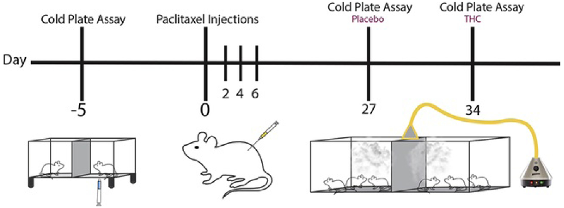
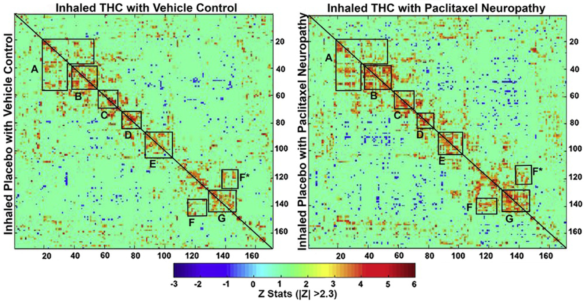
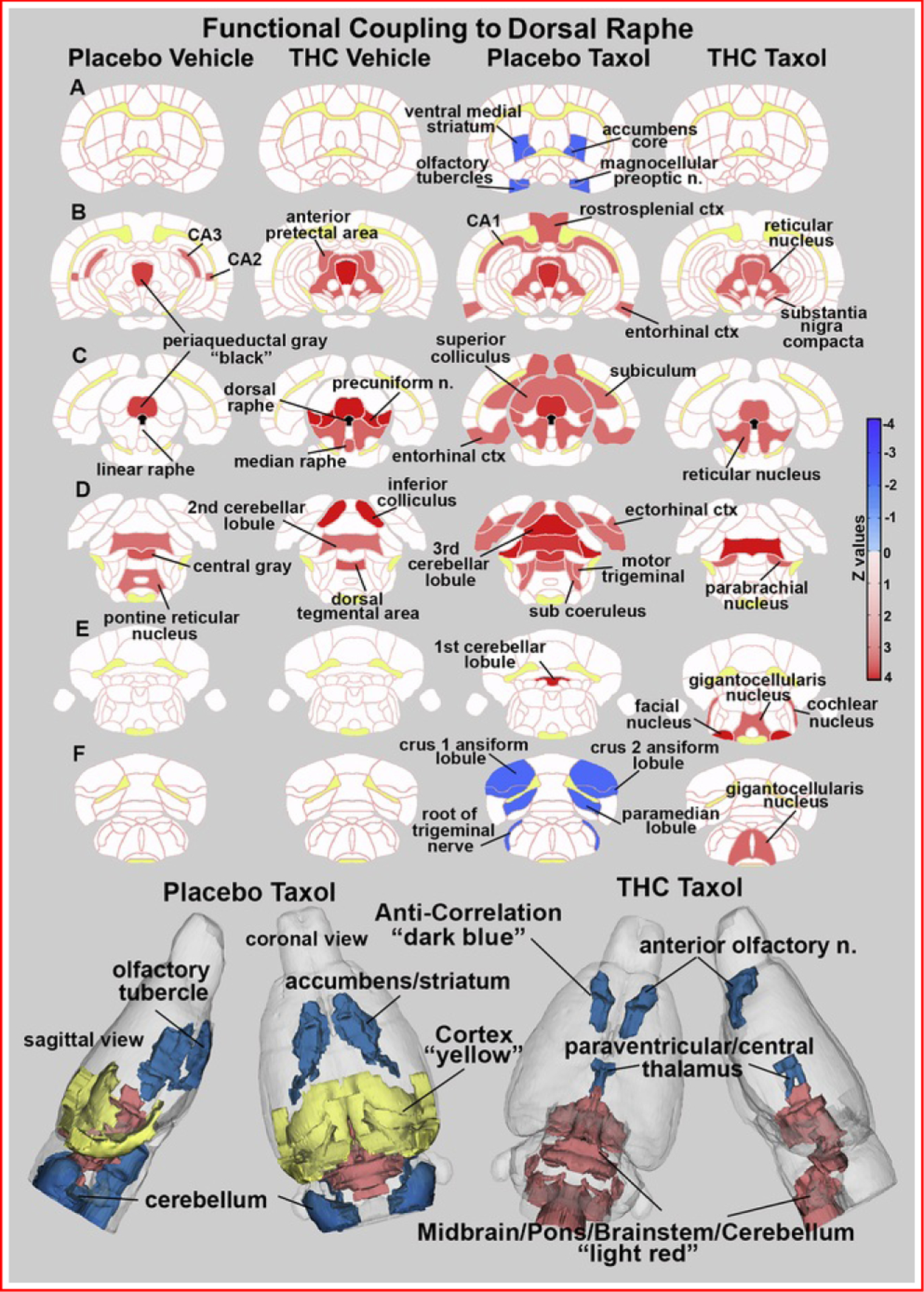
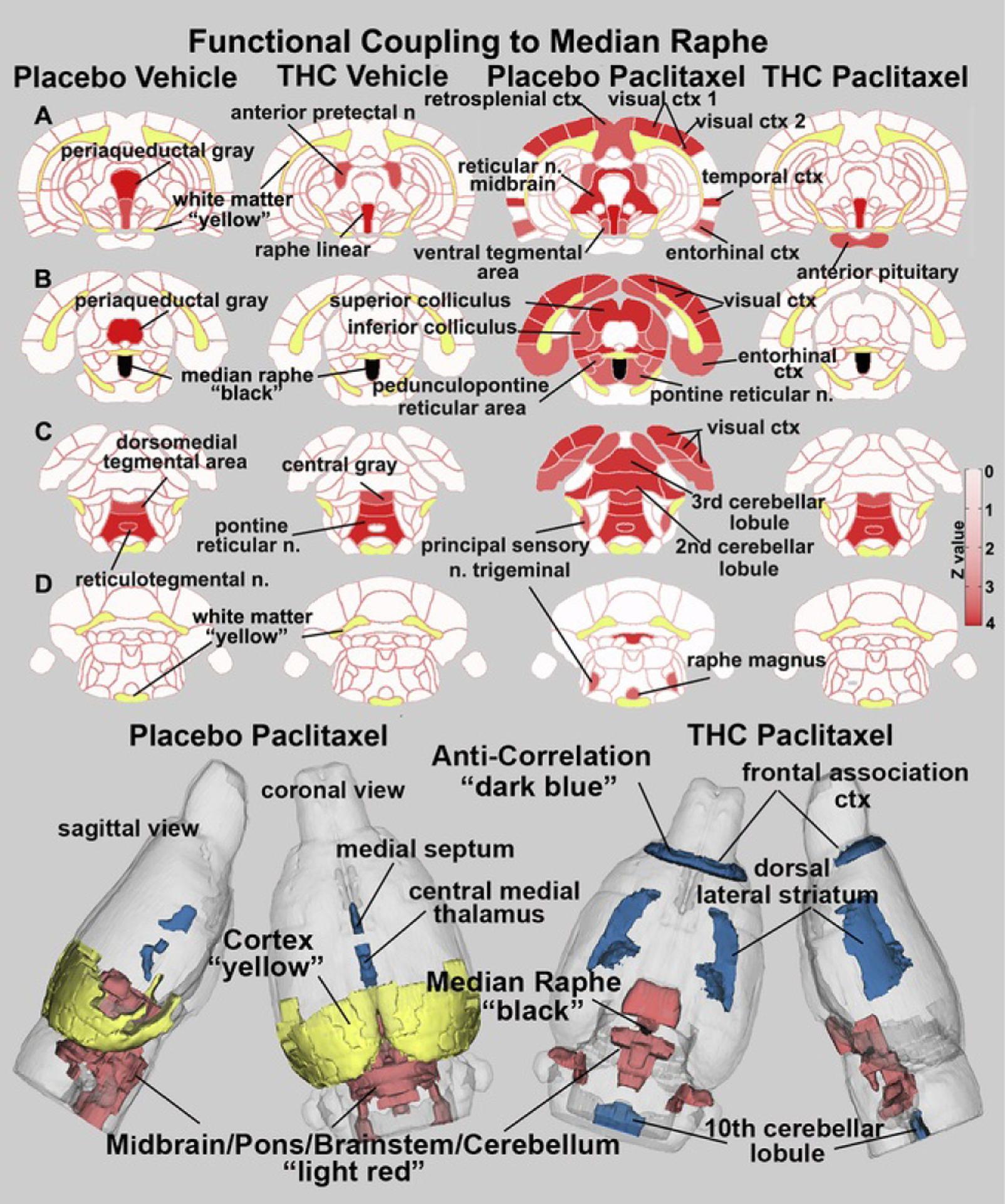
Methods: We assessed the impact of inhalation of vaporized cannabis plant (containing 10.3% Δ9-tetrahydrocannabinol/0.05% cannabidiol) or placebo cannabis on brain resting-state blood oxygen level-dependent functional connectivity and pain behavior induced by paclitaxel in rats. Rats received paclitaxel to produce chemotherapy-induced peripheral neuropathy or its vehicle. Behavioral and imaging experiments were performed after neuropathy was established and stable. Images were registered to, and analyzed using, a 3D magnetic resonance imaging rat atlas providing site-specific data on more than 168 different brain areas.

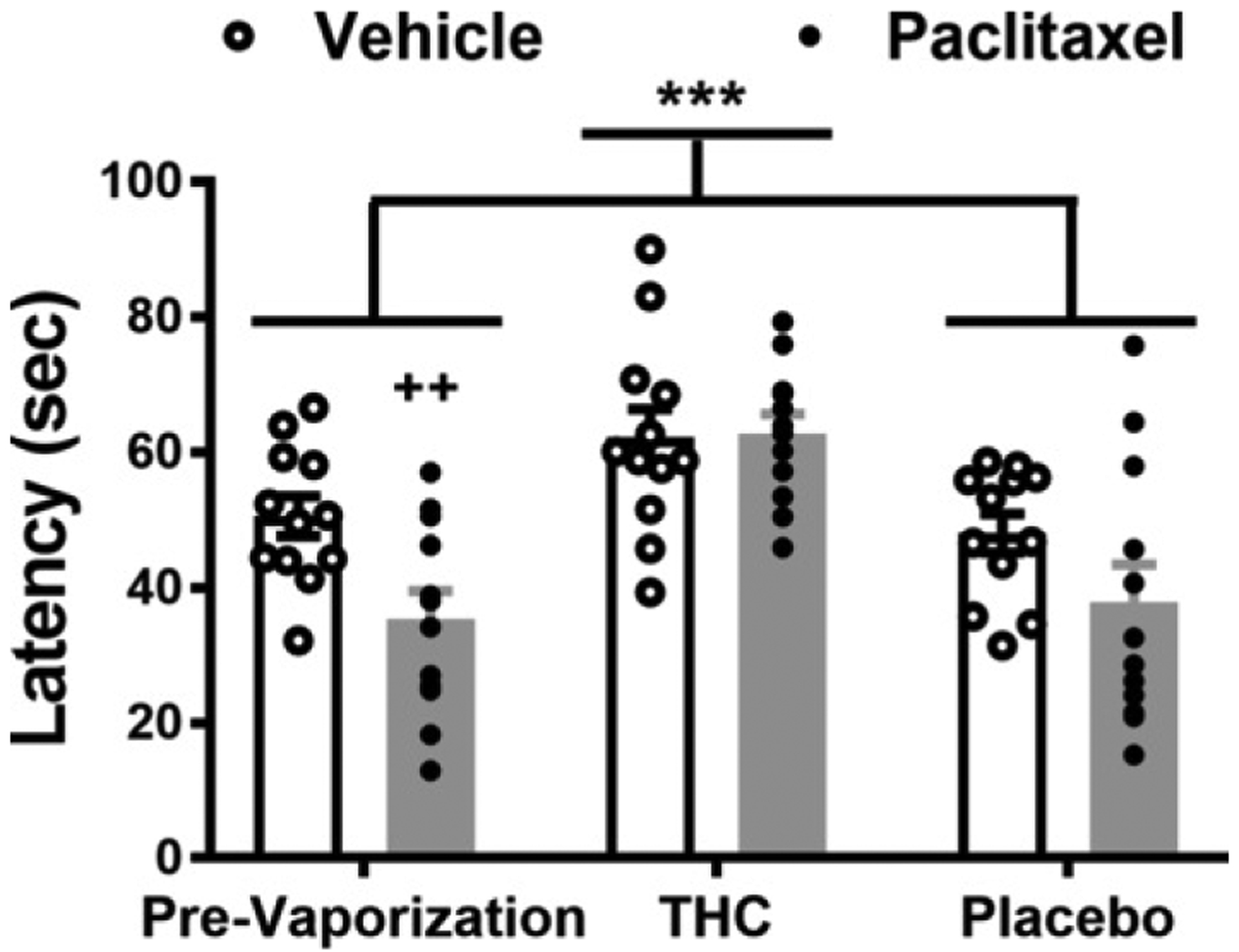
Results: Prior to vaporization, paclitaxel produced cold allodynia. Inhaled vaporized cannabis increased cold withdrawal latencies relative to prevaporization or placebo cannabis, consistent with Δ9-tetrahydrocannabinol-induced antinociception. In paclitaxel-treated rats, the midbrain serotonergic system, comprising the dorsal and median raphe, showed hyperconnectivity to cortical, brainstem, and hippocampal areas, consistent with nociceptive processing. Inhalation of vaporized cannabis uncoupled paclitaxel-induced hyperconnectivity patterns. No such changes in connectivity or cold responsiveness were observed following placebo cannabis vaporization.

Conclusions: Inhaled vaporized cannabis plant uncoupled brain resting-state connectivity in the raphe nuclei, normalizing paclitaxel-induced hyperconnectivity to levels observed in vehicle-treated rats. Inhaled vaporized cannabis produced antinociception in both paclitaxel- and vehicle-treated rats. Our study elucidates neural circuitry implicated in the therapeutic effects of Δ9-tetrahydrocannabinol and supports a role for functional imaging studies in animals in guiding indications for future clinical trials.

Keywords: Cold allodynia; Hyperconnectivity; Paclitaxel; Phytocannabinoids; Resting-state BOLD functional connectivity; Serotonin; Tetrahydrocannabinol; Vaporized marijuana plant.


Conflict of interest statement

Disclosures

CFF has a financial interest in Animal Imaging Research, the company that makes the RF electronics and holders for animal imaging. All other authors report no biomedical financial interests or potential conflicts of interest.

Figures

Figure 1
Figure 1
Experimental timeline. The schematic shows the timeline (days) of experimental steps taken to test for paclitaxel-induced cold allodynia with and without inhalation of vaporized THC cannabis or placebo. THC, Δ9-tetrahydrocannabinol.
Figure 2
Figure 2
Paclitaxel decreases, whereas vaporized THC-enriched cannabis increases, paw withdrawal latencies to cold stimulation. Inhalation of vaporized cannabis containing THC increases paw withdrawal latencies to cold stimulation. Paw withdrawal latencies to cold stimulation were lower in paclitaxel-treated rats compared with vehicle-treated rats, consistent with development of cold allodynia induced by chemotherapy (F1,22 = 5.385, p = .0300). Paw withdrawal latencies were higher following vaporization of THC cannabis compared with placebo cannabis or prevaporization responding irrespective of chemotherapy status (F2,44 = 19.76, p < .0001). Simple-effects analysis revealed that in paclitaxel-treated rats, paw withdrawal latencies were higher following vaporization of THC-enriched cannabis relative to prevaporization levels (p < .0001, Tukey’s multiple comparison test) and vaporization of placebo cannabis (p < .0001, Tukey’s multiple comparison test). In vehicle-treated rats, paw withdrawal latencies were higher following vaporization of THC cannabis compared with placebo cannabis (p = .0203, Tukey’s multiple comparison test) but not compared with prevaporization responding (p = .0659, Tukey’s multiple comparison test). Individual subjects’ responding is shown by the scatter plot. Data are mean ± SEM. ***p < .0001 vs. prevaporization and placebo (two-way [2 × 3] analysis of variance followed by Tukey’s post hoc test). Prior to vaporization, paclitaxel reduced cold withdrawal latencies relative to vehicle treatment (t22 = 3.026, p = .0032 vs. vehicle treatment). ++p < .01, unpaired t test, one tailed. THC, Δ9-tetrahydrocannabinol.
Figure 3
Figure 3
Resting-state functional connectivity. The left correlation matrix compares functional coupling between rats treated with vehicle and later exposed to inhaled vaporized cannabis plant high in THC or placebo. The right matrix compares connectivity between rats treated with paclitaxel to induce cold allodynia and later treated with inhaled placebo or THC. The diagonal line in each matrix separates the two experimental conditions. Each colored red/orange pixel represents 1 of 166 brain areas that has a significant positive correlation with other brain areas. Pixels in shades of blue have a significant negative correlation, or anticorrelation, with other brain regions. The brain areas with significant correlations appear as clusters because they are contiguous in their neuroanatomy and function. Each pixel on one side of the line has a mirror image pixel on the other side. The delineated areas serve to focus attention on similarities and differences in connectivity. Areas: (A) prefrontal cortex (e.g., orbital, prelimbic, infralimbic, and anterior cingulate cortices connections with accumbens, ventral pallidum, basal ganglia, and central amygdala); (B) basal ganglia/hypothalamus/amygdala; (C) thalamus; (D) thalamus/hippocampus; (E) pones/midbrain; (F) cerebellum/brainstem; G, cerebellum. THC, Δ9-tetrahydrocannabinol.
Figure 4
Figure 4
Functional coupling to the dorsal raphe. Paclitaxel produces hyperconnectivity of functional coupling to the dorsal raphe that is attenuated by vaporization of THC-enriched cannabis. The columns of 2D images show heat maps of significant connectivity (z values) for each experimental condition. The shades of red (positive connectivity) and blue (negative connectivity) appear on anatomical sections taken from the rat brain atlas. These sections are identical across rows (A–F). Yellow highlights white matter tracts, and black shows the location of the dorsal raphe in (C). The brain areas with significant connectivity to the dorsal raphe were taken from Table 1. The most rostral brain areas in Table 1 (e.g., anterior olfactory nucleus) are not shown in these 2D sections but are displayed in the 3D color-coded reconstruction of the placebo paclitaxel and THC paclitaxel conditions below. ctx, cortex; n., nucleus; THC, Δ9-tetrahydrocannabinol.
Figure 5
Figure 5
Functional coupling to the median raphe. Paclitaxel produces hyperconnectivity of functional coupling to the median raphe that is attenuated by vaporization of THC-enriched cannabis. The columns of 2D images show heat maps of significant connectivity (z values) for each experimental condition. The shades of red (positive connectivity) appear on anatomical sections taken from the rat brain atlas. These sections are identical across rows (A–D). Yellow highlights white matter tracts, and black shows the location of the median raphe in (B). The brain areas with significant connectivity to the median raphe were taken from Table 2. The most rostral and caudal brain areas showing negative coupling in Table 2 do not appear in the 2D images but are shown in blue in the color-coded 3D reconstruction of placebo paclitaxel and THC paclitaxel conditions below. ctx, cortex; n., nucleus; THC, Δ9-tetrahydrocannabinol.

References

    1. Ibrahim EY and Ehrlich BE, Prevention of chemotherapy-induced peripheral neuropathy: A review of recent findings, Crit Rev Oncol Hematol 145, 2020, 102831. - PMC - PubMed
    1. Seretny M, Currie GL, Sena ES, Ramnarine S, Grant R, MacLeod MR, et al., Incidence, prevalence, and predictors of chemotherapy-induced peripheral neuropathy: A systematic review and meta-analysis, Pain 155,2014, 2461–2470. - PubMed
    1. Szok D, Tajti J, Nyari A and Vecsei L, Therapeutic approaches for peripheral and central neuropathic pain, Behav Neurol 2019, 2019, 8685954. - PMC - PubMed
    1. Smith EM, Pang H, Cirrincione C, Fleishman S, Paskett ED, Ahles T, et al., Effect of duloxetine on pain, function, and quality of life among patients with chemotherapy-induced painful peripheral neuropathy: A randomized clinical trial, JAMA 309, 2013, 1359–1367. - PMC - PubMed
    1. Russo EB, Cannabinoids in the management of difficult to treat pain, Ther Clin Risk Manag 4, 2008, 245–259. - PMC - PubMed

Publication types

Substances