Preconceptual Priming Overrides Susceptibility to Escherichia coli Systemic Infection during Pregnancy
- PMID: 33622714
- PMCID: PMC8545081
- DOI: 10.1128/mBio.00002-21
Preconceptual Priming Overrides Susceptibility to Escherichia coli Systemic Infection during Pregnancy
Abstract
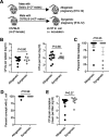
Maternal sepsis is a leading cause of morbidity and mortality during pregnancy. Escherichia coli is a primary cause of bacteremia in women and occurs more frequently during pregnancy. Several key outstanding questions remain regarding how to identify women at highest infection risk and how to boost immunity against E. coli infection during pregnancy. Here, we show that pregnancy-induced susceptibility to E. coli systemic infection extends to rodents as a model of human infection. Mice infected during pregnancy contain >100-fold-more recoverable bacteria in target tissues than nonpregnant controls. Infection leads to near complete fetal wastage that parallels placental plus congenital fetal invasion. Susceptibility in maternal tissues positively correlates with the number of concepti, suggesting important contributions by expanded placental-fetal target tissue. Remarkably, these pregnancy-induced susceptibility phenotypes are also efficiently overturned in mice with resolved sublethal infection prior to pregnancy. Preconceptual infection primes the accumulation of E. coli-specific IgG and IgM antibodies, and adoptive transfer of serum containing these antibodies to naive recipient mice protects against fetal wastage. Together, these results suggest that the lack of E. coli immunity may help discriminate individuals at risk during pregnancy, and that overriding susceptibility to E. coli prenatal infection by preconceptual priming is a potential strategy for boosting immunity in this physiological window of vulnerability.IMPORTANCE Pregnancy makes women especially vulnerable to infection. The most common cause of bloodstream infection during pregnancy is by a bacterium called Escherichia coli This bacterium is a very common cause of bloodstream infection, not just during pregnancy but in all individuals, from newborn babies to the elderly, probably because it is always present in our intestine and can intermittently invade through this mucosal barrier. We first show that pregnancy in animals also makes them more susceptible to E. coli bloodstream infection. This is important because many of the dominant factors likely to control differences in human infection susceptibility can be property controlled for only in animals. Despite this vulnerability induced by pregnancy, we also show that animals with resolved E. coli infection are protected against reinfection during pregnancy, including having resistance to most infection-induced pregnancy complications. Protection against reinfection is mediated by antibodies that can be measured in the blood. This information may help to explain why most women do not develop E. coli infection during pregnancy, enabling new approaches for identifying those at especially high risk of infection and strategies for preventing infection during pregnancy.
Keywords: Escherichia coli; preconceptual; pregnancy; prenatal infection; vaccination.
Copyright © 2021 Prasanphanich et al.
Figures


Similar articles
-
Perinatal Listeria monocytogenes susceptibility despite preconceptual priming and maintenance of pathogen-specific CD8(+) T cells during pregnancy.Cell Mol Immunol. 2014 Nov;11(6):595-605. doi: 10.1038/cmi.2014.84. Epub 2014 Sep 22. Cell Mol Immunol. 2014. PMID: 25242275 Free PMC article.
-
Sex Bias in Sepsis.Cell Host Microbe. 2018 Nov 14;24(5):613-615. doi: 10.1016/j.chom.2018.10.014. Cell Host Microbe. 2018. PMID: 30439335
-
Passive immunity acquisition of maternal anti-enterohemorrhagic Escherichia coli (EHEC) O157:H7 IgG antibodies by the newborn.Eur J Pediatr. 2007 May;166(5):413-9. doi: 10.1007/s00431-006-0250-9. Epub 2006 Oct 21. Eur J Pediatr. 2007. PMID: 17058099
-
Mucosal and systemic responses of immunogenic vaccines candidates against enteric Escherichia coli infections in ruminants: A review.Microb Pathog. 2018 Apr;117:175-183. doi: 10.1016/j.micpath.2018.02.039. Epub 2018 Feb 19. Microb Pathog. 2018. PMID: 29471137 Review.
-
Immune-metabolic adaptations in pregnancy: A potential stepping-stone to sepsis.EBioMedicine. 2022 Dec;86:104337. doi: 10.1016/j.ebiom.2022.104337. Epub 2022 Dec 2. EBioMedicine. 2022. PMID: 36470829 Free PMC article. Review.
Cited by
-
Pilot Study on the Effect of Patient Condition and Clinical Parameters on Hypoxia-Induced Factor Expression: HIF1A, EPAS1 and HIF3A in Human Colostrum Cells.Int J Mol Sci. 2024 Oct 14;25(20):11042. doi: 10.3390/ijms252011042. Int J Mol Sci. 2024. PMID: 39456823 Free PMC article.
-
Maternal bacteremia in intrapartum fever: the role of ampicillin resistance and prolonged membrane rupture-a retrospective comparative study.Arch Gynecol Obstet. 2025 Aug;312(2):451-460. doi: 10.1007/s00404-025-08030-6. Epub 2025 Apr 23. Arch Gynecol Obstet. 2025. PMID: 40266333 Free PMC article.
-
The induction of preterm labor in rhesus macaques is determined by the strength of immune response to intrauterine infection.PLoS Biol. 2021 Sep 8;19(9):e3001385. doi: 10.1371/journal.pbio.3001385. eCollection 2021 Sep. PLoS Biol. 2021. PMID: 34495952 Free PMC article.
-
Antimicrobial Resistance in Maternal Infections During Pregnancy.Biomedicines. 2025 Mar 22;13(4):777. doi: 10.3390/biomedicines13040777. Biomedicines. 2025. PMID: 40299356 Free PMC article.
-
The right educational environment: Oral tolerance in early life.Immunol Rev. 2024 Sep;326(1):17-34. doi: 10.1111/imr.13366. Epub 2024 Jul 13. Immunol Rev. 2024. PMID: 39001685 Free PMC article. Review.
References
Publication types
MeSH terms
Substances
Grants and funding
LinkOut - more resources
Full Text Sources
Other Literature Sources
Medical
