Molecular Basis of the Versatile Regulatory Mechanism of HtrA-Type Protease AlgW from Pseudomonas aeruginosa
- PMID: 33622718
- PMCID: PMC8545111
- DOI: 10.1128/mBio.03299-20
Molecular Basis of the Versatile Regulatory Mechanism of HtrA-Type Protease AlgW from Pseudomonas aeruginosa
Erratum in
-
Erratum for Li et al., "Molecular Basis of the Versatile Regulatory Mechanism of HtrA-Type Protease AlgW from Pseudomonas aeruginosa".mBio. 2021 Aug 31;12(4):e0164021. doi: 10.1128/mBio.01640-21. Epub 2021 Jul 13. mBio. 2021. PMID: 34253068 Free PMC article. No abstract available.
Abstract
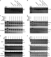
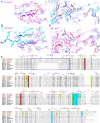
AlgW, a membrane-bound periplasmic serine protease belonging to the HtrA protein family, is a key regulator of the regulated intramembrane proteolysis (RIP) pathway and is responsible for transmitting the envelope stress signals in Pseudomonas aeruginosa The AlgW PDZ domain senses and binds the C-terminal of mis-localized outer membrane proteins (OMPs) or periplasmic protein MucE, leading to catalytic activation of the protease domain. While AlgW is functionally well studied, its exact activation mechanism remains to be elucidated. Here, we show that AlgW is a novel HtrA protease that can be biochemically activated by both peptide and lipid signals. Compared with the corresponding homologue DegS in Escherichia coli, AlgW exhibits a distinct substrate specificity and regulation mechanism. Structural, biochemical, and mutagenic analyses revealed that, by specifically binding to the C-terminal decapeptide of MucE, AlgW could adopt more relaxed conformation and obtain higher activity than with tripeptide activation. We also investigated the regulatory mechanism of the LA loop in AlgW and proved that the unique structural feature of this region was responsible for the distinct enzymatic property of AlgW. These results demonstrate the unique and diverse activation mechanism of AlgW, which P. aeruginosa may utilize to enhance its adaptability to environmental stress.IMPORTANCE HtrA-family proteases are commonly employed to sense the protein folding stress and activate the regulated intramembrane proteolysis (RIP) cascade in Gram-negative bacteria. Here, we reveal the unique dual-signal activation and dynamic regulation properties of AlgW, an HtrA-type protease triggering the AlgU stress-response pathway, which controls alginate production and mucoid conversion in Pseudomonas aeruginosa The structural and functional data offer insights into the molecular basis underlying the transition of different activation states of AlgW in response to different effectors. Probing these unique features provides an opportunity to correlate the diverse regulation mechanism of AlgW with the high adaptability of P. aeruginosa to environmental changes during infection.
Keywords: AlgW; HtrA; Pseudomonas aeruginosa; alginate; crystal structure; mucoid phenotype; regulated intramembrane proteolysis.
Copyright © 2021 Li et al.
Figures


Similar articles
-
Two distinct loci affecting conversion to mucoidy in Pseudomonas aeruginosa in cystic fibrosis encode homologs of the serine protease HtrA.J Bacteriol. 1996 Jan;178(2):511-23. doi: 10.1128/jb.178.2.511-523.1996. J Bacteriol. 1996. PMID: 8550474 Free PMC article.
-
Pseudomonas aeruginosa Regulated Intramembrane Proteolysis: Protease MucP Can Overcome Mutations in the AlgO Periplasmic Protease To Restore Alginate Production in Nonmucoid Revertants.J Bacteriol. 2018 Jul 25;200(16):e00215-18. doi: 10.1128/JB.00215-18. Print 2018 Aug 15. J Bacteriol. 2018. PMID: 29784885 Free PMC article.
-
Cell wall-inhibitory antibiotics activate the alginate biosynthesis operon in Pseudomonas aeruginosa: Roles of sigma (AlgT) and the AlgW and Prc proteases.Mol Microbiol. 2006 Oct;62(2):412-26. doi: 10.1111/j.1365-2958.2006.05390.x. Mol Microbiol. 2006. PMID: 17020580
-
Function, molecular mechanisms, and therapeutic potential of bacterial HtrA proteins: An evolving view.Comput Struct Biotechnol J. 2021 Dec 8;20:40-49. doi: 10.1016/j.csbj.2021.12.004. eCollection 2022. Comput Struct Biotechnol J. 2021. PMID: 34976310 Free PMC article. Review.
-
Regulated proteolysis: control of the Escherichia coli σ(E)-dependent cell envelope stress response.Subcell Biochem. 2013;66:129-60. doi: 10.1007/978-94-007-5940-4_6. Subcell Biochem. 2013. PMID: 23479440 Review.
Cited by
-
HtrAs are essential for the survival of the haloarchaeon Natrinema gari J7-2 in response to heat, high salinity, and toxic substances.Appl Environ Microbiol. 2024 Feb 21;90(2):e0204823. doi: 10.1128/aem.02048-23. Epub 2024 Jan 30. Appl Environ Microbiol. 2024. PMID: 38289131 Free PMC article.
-
Molecular mechanism of the one-component regulator RccR on bacterial metabolism and virulence.Nucleic Acids Res. 2024 Apr 12;52(6):3433-3449. doi: 10.1093/nar/gkae171. Nucleic Acids Res. 2024. PMID: 38477394 Free PMC article.
-
Environmental alkalization suppresses deployment of virulence strategies in Pseudomonas syringae pv. tomato DC3000.J Bacteriol. 2024 Nov 21;206(11):e0008624. doi: 10.1128/jb.00086-24. Epub 2024 Oct 24. J Bacteriol. 2024. PMID: 39445803 Free PMC article.
-
Study of andrographolide bioactivity against Pseudomonas aeruginosa based on computational methodology and biochemical analysis.Front Chem. 2024 Apr 12;12:1388545. doi: 10.3389/fchem.2024.1388545. eCollection 2024. Front Chem. 2024. PMID: 38680458 Free PMC article.
-
The PDZ domain of EpsC is required for extracellular secretion of VesB by the Type II secretion system in Vibrio cholerae.J Bacteriol. 2025 Aug 21;207(8):e0014425. doi: 10.1128/jb.00144-25. Epub 2025 Jul 14. J Bacteriol. 2025. PMID: 40657929 Free PMC article.
References
-
- Chevalier S, Bouffartigues E, Bazire A, Tahrioui A, Duchesne R, Tortuel D, Maillot O, Clamens T, Orange N, Feuilloley MGJ, Lesouhaitier O, Dufour A, Cornelis P. 2019. Extracytoplasmic function sigma factors in Pseudomonas aeruginosa. Biochim Biophys Acta Gene Regul Mech 1862:706–721. doi:10.1016/j.bbagrm.2018.04.008. - DOI - PubMed
Publication types
MeSH terms
Substances
LinkOut - more resources
Full Text Sources
Other Literature Sources
Miscellaneous

